Thoracostomy
A thoracostomy is a small incision of the chest wall, with maintenance of the opening for drainage.[1] It is most commonly used for the treatment of a pneumothorax. This is performed by physicians, emergency response nurses, and paramedics, usually via needle thoracostomy or with a thoracostomy tube (chest tube).
| Thoracostomy | |
|---|---|
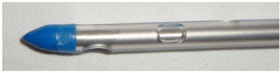 A tube thoracostomy unit | |
| Specialty | Pulmonology |
| ICD-10-PCS | Z46.82 |
| MeSH | D006468 |
| MedlinePlus | 002947 |
| eMedicine | 80678 |
A thoracostomy is often confused with thoracotomy, which is a larger incision commonly used to gain access to organs within the chest.
Medical uses
When air, blood, or other fluids accumulate in the pleural cavity it may be drained by thoracostomy. Whereas air in this space (pneumothorax) may be released by needle thoracostomy, other substances require drainage with a thoracostomy tube.[2]
Contra-indications
There are no absolute contraindications to thoracostomy. There are relative contraindications (such as coagulopathies); however, in an emergency setting these are outweighed by the necessity to re-inflate a collapsed lung by draining fluid/air from the space around the lung.[2]
Technique
Drainage of the pleural cavity is achieved by the surgeon making a primary incision in the skin followed by a second incision through the muscle between the ribs. This way a tube may be guided into the chest to allow for drainage. Chest tubes are designed to collect this drainage and prevent anything from leaking back into the pleural space. This is accomplished by a check valve, usually part of a specialized drainage system with an underwater seal. Depending on the amount of air/fluid to be drained, the collection bottle may need to be periodically changed.[2]
Risks/complications
Rare complications are mostly due to placement technique, inexperience of the interventionist, and emergent vs. elective circumstances. The most common complications are recurrent pneumothorax (incomplete recovery, but an expected course), infection, and organ injury (due to mechanical damage).[3]
Esophageal injury is rare. If saliva and chyme contents drain from the chest tube, that should raise suspicion of esophageal injury. The main treatment of esophageal injury is surgical repair. The stomach is also rarely injured. Proper technique and not using a trocar during the procedure decreases the risk of this from occurring. [3]
See also
References
- Dorland, W. A. Newman (2009). Dorland's pocket medical dictionary (28th ed.). Philadelphia, PA: Saunders/Elsevier. ISBN 978-1-4160-3420-9.
- Nicks, Bret A.; Manthey, David (2011). Pneumothorax. Tintinalli's Emergency Medicine. New York City: McGraw-Hill.
- Kwiatt, Michael; Tarbox, Abigail; Seamon, Mark J.; Swaroop, Mamta; Cipolla, James; Allen, Charles; Hallenbeck, Stacinoel; Davido, H. Tracy; Lindsey, David E. (2014). "Thoracostomy tubes: A comprehensive review of complications and related topics". International Journal of Critical Illness and Injury Science. 4 (2): 143–155. doi:10.4103/2229-5151.134182. ISSN 2229-5151. PMC 4093965. PMID 25024942.