Superficial peroneal nerve
The superficial peroneal nerve innervates the peroneus longus and peroneus brevis muscles and the skin over the antero-lateral aspect of the leg along with the greater part of the dorsum of the foot (with the exception of the first web space, which is innervated by the deep peroneal nerve).
| Superficial peroneal nerve | |
|---|---|
 Deep nerves of the front of the leg. | |
| Details | |
| From | Common peroneal nerve |
| To | Medial dorsal cutaneous nerve, intermediate dorsal cutaneous nerve |
| Identifiers | |
| Latin | Nervus fibularis superficialis, nervus peronaeus superficialis |
| TA | A14.2.07.050 |
| FMA | 44699 |
| Anatomical terms of neuroanatomy | |
Structure
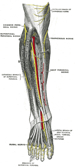
Lateral side of the leg
Superficial peroneal nerve is the main nerve of the lateral compartment of the leg. It begins at the lateral side of the neck of fibula, and runs through the peroneal muscles. In the middle third of the leg, it descends between the peroneus longus and peroneus brevis muscles, and then reaches the anterior border of the peroneus brevis to enter the groove between the peroneus brevis and extensor digitorum longus under the deep fascia of leg. It becomes superficial at the junction of upper two-thirds and lower one-thirds of the leg by piercing the deep fascia. Superficial peroneal nerve gives off several branches in the leg.[1]
- Muscular branches to peroneus longus and peroneus brevis[1]
- Cutaneous branches to supplies the skin over the lower one-third of the lateral side of the leg and greater part of the dorsum of the foot except for areas that are supplied by saphenous nerve (medial side of the leg), sural nerve (lateral side of the foot), deep peroneal nerve (first webbed space of the dorsum of the foot), medial and lateral plantar nerves (plantar surface of the foot).[1]
Foot
At the junction between the upper two-thirds and lower one-thirds of the leg, superficial peroneal nerve is divided into medial dorsal cutaneous nerve (medial branch) and intermediate dorsal cutaneous nerve (lateral branch).[1]
- The medial branch crosses the ankle and divides into two dorsal digital nerves - one for the medial side of the big toe, and the other for the adjoining sides of the 2nd and 3rd toes.[1]
- The lateral branch divides into two dorsal digital nerves for the adjoining sides of the third and fourth, and fourth and fifth toes.[1]
- Communicating branches - the medial branch communicates with saphenous nerve and deep peroneal nerves while the lateral branch communicates with sural nerve.[1]
Clinical significance
Injury to the nerve can result in an inability to evert the foot and loss of sensation over the dorsum of the foot (with the exception of the first web space between the great toe and the second toe, where the deep fibular nerve innervates).
Additional images
 Cutaneous nerves of the right lower extremity. Front and posterior views.
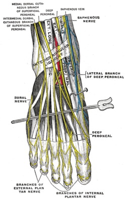 Nerves of the dorsum of the foot.
References
- Krishna, Garg (2010). "Front, lateral, and medial sides of leg and dorsum of foot (Chapter 8)". BD Chaurasia's Human Anatomy (Regional and Applied Dissection and Clinical) Volume 2 - Lower limb, abdomen, and pelvis (Fifth ed.). India: CBS Publishers and Distributors Pvt Ltd. p. 109,110. ISBN 978-81-239-1864-8.
External links
- Anatomy photo:15:st-0505 at the SUNY Downstate Medical Center - "The Leg - Nerves"
| Authority control |
|---|