Subependymoma
A subependymoma is a type of brain tumor; specifically, it is a rare form of ependymal tumor.[1]They are usually in middle aged people. Earlier, they were called subependymal astrocytomas.[2]
| Subependymoma | |
|---|---|
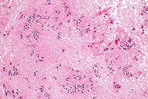 | |
| Micrograph of a subependymoma showing the characteristic clustering of nuclei. H&E stain. | |
| Specialty | Neoplasms |
The prognosis for a subependymoma is better than for most ependymal tumors,[3] and is considered a grade I tumor in the World Health Organization (WHO) classification.
They are classically found within the fourth ventricle, typically have a well demarcated interface to normal tissue and do not usually extend into the brain parenchyma, like ependymomas often do.[4]
Symptoms
Patients are often asymptomatic, and are incidentally diagnosed. Larger tumours are often with increased intracranial pressure.[2]
Pathology
These tumours are small, no more than two centimeters across, coming from the ependyma. The best way to distinguish it from a subependymal giant cell astrocytoma is the size.[2]
Diagnosis
The diagnosis is based on tissue, e.g. a biopsy. Histologically subependymomas consistent of microcystic spaces and bland appearing cells without appreciable nuclear atypia or mitoses. The nuclei tend to form clusters.
On a CT, it often shows a less dense to equalle dense mass. If it is big, it may have parts that are cystic or calcific.[2]In 50-60% of cases, the tumor is in the fourth ventricle, while the second most common (30-40% of cases) location is the side ventricles. It is rare for it to be in the third ventricle or the central canal of the spinal cord.[2]
Treatment
Asymptomatic cases may only need watchful waiting. If symptomatic, it can be surgically removed, and partial removal also carries an excellent prognosis.[2]
Prognosis
The outlook of a cure is extremely favorable.[2]
References
- Orakcioglu B, Schramm P, Kohlhof P, Aschoff A, Unterberg A, Halatsch ME (January 2009). "Characteristics of thoracolumbar intramedullary subependymomas". J Neurosurg Spine. 10 (1): 54–9. doi:10.3171/2008.10.SPI08311. PMID 19119934.
- Gaillard, Frank. "Subependymoma | Radiology Reference Article | Radiopaedia.org". radiopaedia.org. Retrieved 2018-04-15.
- Prayson RA, Suh JH (April 1999). "Subependymomas: clinicopathologic study of 14 tumors, including comparative MIB-1 immunohistochemical analysis with other ependymal neoplasms". Arch. Pathol. Lab. Med. 123 (4): 306–9. doi:10.1043/0003-9985(1999)123<0306:S>2.0.CO;2 (inactive 2019-08-19). PMID 10320142.
- Hoeffel, C.; Boukobza, M.; Polivka, M.; Lot, G.; Guichard, JP.; Lafitte, F.; Reizine, D.; Merland, JJ. (1995). "MR manifestations of subependymomas". AJNR Am J Neuroradiol. 16 (10): 2121–9. PMID 8585504.
External links
| Classification |
|
|---|