Subdural hygroma
A subdural hygroma is a collection of cerebrospinal fluid (CSF), without blood, located under the dural membrane. Most subdural hygromas are believed to be derived from chronic subdural hematomas. They are commonly seen in elderly people after minor trauma but can also be seen in children after an infection. One of the common causes of subdural hygroma is a sudden decrease in pressure as a result of placing a ventricular shunt. This can lead to leakage of CSF into the subdural space especially in cases with moderate to severe brain atrophy. In these cases the symptoms such as mild fever, headache, drowsiness and confusion can be seen, which are relieved by draining this subdural fluid.
| Subdural hygroma | |
|---|---|
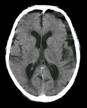 | |
| Subdural hygroma, frontal and temporal. Man of 80 years old. | |
| Specialty | Neurology |
Signs and symptoms
Most subdural hygromas are small and clinically insignificant. Larger hygromas may cause secondary localized mass effects on the adjacent brain parenchyma, enough to cause a neurologic deficit or other symptoms. Acute subdural hygromas can be a potential neurosurgical emergency, requiring decompression. Acute hygromas are typically a result of head trauma—they are a relatively common posttraumatic lesion—but can also develop following neurosurgical procedures, and have also been associated with a variety of conditions, including dehydration in the elderly, lymphoma and connective tissue diseases.
Diagnosis
In the majority of cases, if there has not been any acute trauma or severe neurologic symptoms, a small subdural hygroma on the head CT scan will be an incidental finding. If there is an associated localized mass effect that may explain the clinical symptoms, or concern for a potential chronic SDH that could rebleed, then an MRI, with or without neurologic consultation, may be useful.
It is not uncommon for chronic subdural hematomas (SDHs) on CT reports for scans of the head to be misinterpreted as subdural hygromas, and vice versa. Magnetic resonance imaging (MRI) should be done to differentiate a chronic SDH from a subdural hygroma, when clinically warranted. Elderly patients with marked cerebral atrophy, and secondary widened subarachnoid CSF spaces, can also cause confusion on CT. To distinguish chronic subdural hygromas from simple brain atrophy and CSF space expansion, a gadolinium-enhanced MRI can be performed. Visualization of cortical veins traversing the collection favors a widened subarachnoid space as seen in brain atrophy, whereas subdural hygromas will displace the cortex and cortical veins.
Treatment
Most subdural hygromas that are asymptomatic do not require any treatment. Some might opt to perform a simple burr-holes to alleviate intracranial pressure (ICP). Occasionally a temporary drain is placed for 24-48 hours post op. In recurrent cases a craniotomy may be performed to attempt to locate the location of the CSF Leak. In certain cases a shunt can be placed for additional drainage. Great caution is used when choosing to look for the CSF leak due to them generally being difficult to spot.
References
- Taveras, Juan M. et al., eds. Radiology: Diagnosis, Imaging, Intervention. 1994- ISBN 0-397-57115-1; ch. 37: 9-13.
- Brain Inj 1998 Jul;12(7):595-603.
- McCluney KW, Yeakley JW, Fenstermacher MJ, et al. «Subdural hygroma versus atrophy on MR brain scans: "the cortical vein sign"». AJNR Am J Neuroradiol 1992;13: 1335–39.
External links
https://thejns.org/focus/view/journals/neurosurg-focus/26/6/article-pE8.xml
| Classification |
|---|