Stippling (dentistry)
The gingiva often possess a textured surface that is referred to as being stippled (engraved points).[1] Stippling only presents on the attached gingiva bound to underlying alveolar bone, not the freely moveable alveolar mucosa. Stippling used to be thought to indicate health, but it has since been shown that smooth gingiva is not an indication of disease, unless it is smooth due to a loss of previously existing stippling.
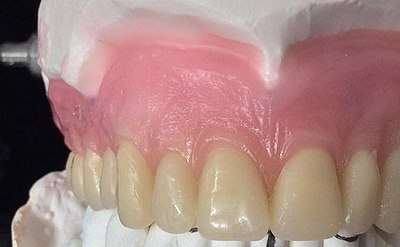
Stippling is a consequence of the microscopic elevations and depressions of the surface of the gingival tissue due to the connective tissue projections within the tissue.[1] "The degree of keratinization and the prominence of stippling appear to be related."[1] To be more specific, stippling occurs at sites of fusion of the epithelial ridges (also known as rete pegs) and correspond to the fusion of the valleys created by the connective tissue papillae.[2]
References
- Itoiz, ME; Carranza, FA: The Gingiva. In Newman, MG; Takei, HH; Carranza, FA; editors: Carranza’s Clinical Periodontology, 9th Edition. Philadelphia: W.B. Saunders Company, 2002. page 30.
- Lindhe's Clinical Periodontology and Implant Dentistry, 4th Ed.