Sternoclavicular joint
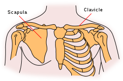
The sternoclavicular joint or sternoclavicular articulation is the joint between the manubrium of the sternum and the clavicle bone. It is structurally classed as a synovial saddle joint and functionally classed as a diarthrosis and multiaxial joint. It is composed of two portions separated by an articular disc of fibrocartilage. The bone areas entering into its formation are the sternal end of the clavicle, the upper and lateral part of the sternum, (the clavicular notch), and the cartilage of the first rib, visible from the outside as the suprasternal notch. The articular surface of the clavicle is much larger than that of the sternum, and is invested with a layer of cartilage, which is considerably thicker than that on the sternum.
| Sternoclavicular joint | |
|---|---|
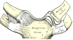 Sternoclavicular joint. Anterior view. | |
 Sternoclavicular joint visible near center but not labeled. | |
| Details | |
| Identifiers | |
| Latin | articulatio sternoclavicularis |
| MeSH | D013247 |
| TA | A03.5.04.001 |
| FMA | 25883 |
| Anatomical terminology | |
The costoclavicular ligament is the main limitation to movement, and therefore the main stabilizer of the joint. A fibrocartilaginous disc present at the joint increases the range of movement. Sternoclavicular dislocation is rare,[1] but may result from direct trauma to the clavicle or indirect forces applied to the shoulder.[2] Posterior dislocations deserve special attention, as they have the potential to be life-threatening because of the risk of damage to vital structures in the mediastinum.[3]
Structure
Function
The sternoclavicular joint allows movement of the clavicle in three planes, predominantly in the anteroposterior and vertical planes, although some rotation also occurs. A description of movement would be elevation and depression. Muscles don't directly act on this joint, although almost all actions of the shoulder girdle or the scapula will cause some motion at this articulation.
The unique double-hinged articular disk found at the junction of the clavicular head and manubrium allows for movement between the clavicle and the disk during elevation and depression of the scapula. This disk also allows motion between the sternum (manubrium) and itself during protraction and retraction of the scapula.[4]
Clinical significance
Trauma may result in the dislocation of the sternoclavicular joint. Posterior dislocation puts the mediastinal structures at risk. A spontaneous partial dislocation can also occur sometimes. In SAPHO syndrome there may be arthropathy of the sternoclavicular joint. Septic arthritis may rarely affect the sternoclavicular joint.
See also
- Acromioclavicular joint
- Shoulder
- Shoulder girdle (Pectoral girdle)
- Shoulder joint
References
- Cadogan, Mike (February 2010). "Sternoclavicular Joint Dislocations". Life in the Fast Lane. Retrieved June 2011. Check date values in:
|accessdate=(help) - Arend CF. Ultrasound of the Shoulder. Master Medical Books, 2013. Free section on sternoclavicular joint dislocation available at ShoulderUS.com
- Jougon, Jacques B.; Lepront, Denis J.; Dromer, Claire E.H. (1996). "Posterior dislocation of the sternoclavicular joint leading to mediastinal compression". The Annals of Thoracic Surgery. 61 (2): 711–3. doi:10.1016/0003-4975(95)00745-8. PMID 8572795.
- Lippert, Lynn. Clinical Kinesiology and Anatomy, 4th edition; pg.95-96.
- This article incorporates text in the public domain from page 313 of the 20th edition of Gray's Anatomy (1918)
External links
| Wikimedia Commons has media related to Sternoclavicular joint. |
- Overview at ouhsc.edu
- Anatomy figure: 10:01-08 at Human Anatomy Online, SUNY Downstate Medical Center
| Authority control |
|---|