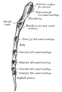
Sternal angle
The sternal angle (also known as the angle of Louis or manubriosternal junction) is the synarthrotic joint formed by the articulation of the manubrium and the body of the sternum.[1][2]
| Sternal angle | |
|---|---|
 Lateral border of sternum | |
 Anterior surface of sternum and costal cartilages. (Sternal angle not labeled, but visible at second costal cartilage.) | |
| Details | |
| Identifiers | |
| Latin | angulus sterni, angulus sternalis, angulus Ludovici |
| TA | A02.3.03.005 |
| FMA | 7547 |
| Anatomical terminology | |
The sternal angle is a palpable clinical landmark in surface anatomy.
Anatomy
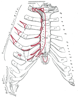
The sternal angle, which varies around 162 degrees in males,[3] marks the approximate level of the 2nd pair of costal cartilages, which attach to the second ribs, and the level of the intervertebral disc between T4 and T5.[2] In clinical applications, the sternal angle can be palpated at the T4 vertebral level.
The sternal angle is used in the definition of the thoracic plane. This marks the level of a number of other anatomical structures:
Structures at the level of the thoracic plane |
|---|
|
- Position of the deep cardiac plexus
- Junction of the intra and extra pericardial parts of the superior vena cava.
- Line junction between C4 and T2 dermatome.
The angle also marks a number of other features:
- Carina of the trachea is deep to the sternal angle
- Passage of the thoracic duct from right to left behind esophagus
- Ligamentum arteriosum
- Loop of left recurrent laryngeal nerve around aortic arch
The angle is in the form of a secondary cartilaginous joint (symphysis).
This is where the 2nd rib joins with the sternum. A clinically useful feature of the (manubriosternal) joint is that it can be palpated easily. This is because the manubrium normally angles posteriorly on the body of the sternum, forming a raised feature referred to as the sternal angle.
History
The sternal angle is also called the angle of Louis, but the reason for that name was lost. Once thought to be after Antoine Louis or Wilhelm Friedrich von Ludwig, it is now believed to be after Pierre Charles Alexandre Louis.[6]
See also
References
- Dalley, Arthur F.; Moore, Keith L. Clinically Oriented Anatomy. Hagerstown, MD: Lippincott Williams & Wilkins. ISBN 0-7817-5936-6.
- Wilson, Herbert H. Srebnik ; illustrations by Genevieve M. (2002). Concepts in anatomy. Boston: Kluwer Academic Publishers. p. 70. ISBN 0792375394.
- Susan Standring; Neil R. Borley; et al., eds. (2008). "Chapter 54: Chest wall and breast". Gray's anatomy : the anatomical basis of clinical practice (40th ed.). London: Churchill Livingstone. p. 922. ISBN 978-0-8089-2371-8.
- Arai et al. Radiographic landmarks of the upper margin of the superior vena cava (SVC) in children Canadian Journal of Anesthesia 49 (Supplement 1): 32
- Viscera of the Thorax UAMS Department of Anatomy
- Alberto Coscione, L. Dixon, H. Ellis (2013). "The "Angle of Louis"" (PDF). Eur. J. Anat. 17 (3): 190–192.CS1 maint: multiple names: authors list (link)
External links
- Anatomy photo:18:st-0212 at the SUNY Downstate Medical Center - "Thoracic Wall: Bones"
| Authority control |
|---|