Cervical cancer staging
Cervical cancer staging is the assessment of cervical cancer to decide how far the disease has progressed. Cancer staging generally runs from stage 0, which is pre-cancerous or non-invasive, to stage IV, in which the cancer has spread throughout a significant part of the body.[1]
Cervical cancer is staged by the International Federation of Gynecology and Obstetrics (FIGO) staging system, which is based on clinical examination, rather than surgical findings.[2] It allows only the following diagnostic tests to be used in determining the stage: palpation (feeling with the fingers), inspection, colposcopy, endocervical curettage, hysteroscopy, cystoscopy, proctoscopy, intravenous urography, and X-ray examination of the lungs and skeleton, and cervical conization.
Stages
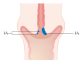 Stage 1A cervical cancer
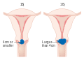 Stage 1B cervical cancer
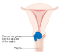 Stage 2A cervical cancer
 Stage 2B cervical cancer
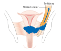 Stage 3B cervical cancer
 Stage 4A cervical cancer
 Stage 4B cervical cancer
- Stage 0
- The carcinoma is confined to the surface layer (cells lining) of the cervix. Also called carcinoma in situ (CIS).
- Stage I
- The carcinoma has grown deeper into the cervix, but has not spread beyond it (extension to the corpus would be disregarded). Stage One is subdivided as follows:
- IA Invasive carcinoma which can be diagnosed only by microscopy, with deepest invasion ≤5 mm and the largest extension ≤7 mm
- IA-1 Measured stromal invasion of ≤3.0 mm in depth and extension of ≤7.0 mm
- IA-2 Measured stromal invasion of >3.0 mm and not >5.0 mm with an extension of not >7.0 mm
- IB Clinically visible lesions limited to the cervix uteri or pre-clinical cancers greater than stage IA
- IB-1 Clinically visible lesion <4.0 cm in greatest dimension
- IB-2 Clinically visible lesion >4.0 cm in greatest dimension
- IA Invasive carcinoma which can be diagnosed only by microscopy, with deepest invasion ≤5 mm and the largest extension ≤7 mm
- Stage II
- Cervical carcinoma invades beyond the uterus, but not to the pelvic wall or to the lower third of the vagina
- IIA Without parametrial invasion
- IIA-1 Clinically visible lesion <4.0 cm in greatest dimension
- IIA-2 Clinically visible lesion >4.0 cm in greatest dimension
- IIB With obvious parametrial invasion
- IIA Without parametrial invasion
- Stage III
- The tumour extends to the pelvic wall and/or involves lower third of the vagina and/or causes hydronephrosis or non-functioning kidney
- IIIA Tumour involves lower third of the vagina, with no extension to the pelvic wall
- IIIB Extension to the pelvic wall and/or hydronephrosis or non-functioning kidney
- Stage IV
- The carcinoma has extended beyond the true pelvis or has involved (biopsy proven) the mucosa of the bladder or rectum. A bullous oedema, as such, does not permit a case to be allotted to Stage IV
- IVA Spread of the growth to adjacent organs
- IVB Spread to distant organs[3]
References
- "Staging". National Cancer Institute. Retrieved 2018-11-07.
- "Cervical Cancer Stages". www.cancer.org. Retrieved 2018-11-07.
- Rosdahl, Caroline (2012). Textbook of basic nursing. Philadelphia: Wolters Kluwer Health/Lippincott Williams & Wilkins. p. 1550. ISBN 1605477729.