Somatopleuric mesenchyme
In the anatomy of an embryo, the somatopleuric mesenchyme is a structure created during embryogenesis when the lateral mesoderm splits into two layers. The outer (or somatic) layer becomes applied to the inner surface of the ectoderm, and with it forms the somatopleure.
| Somatopleuric mesenchyme | |
|---|---|
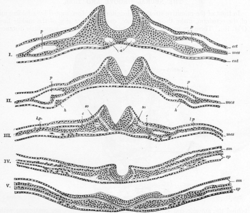 A series of a transverse sections through an embryo of the dog. (After Bonnet.) Section I is the most anterior. In V the neural plate is spread out nearly flat. The series shows the uprising of the neural folds to form the neural canal. a. Aortæ. c. Intermediate cell mass. ect. Ectoderm. ent. Entoderm. h, h. Rudiments of endothelial heart tubes. In III, IV, and V the scattered cells represented between the entoderm and splanchnic layer of mesoderm are the vasoformative cells which give origin in front, according to Bonnet, to the heart tubes, h; l.p. Lateral plate still undivided in I, II, and III; in IV and V split into somatic (sm) and splanchnic (sp) layers of mesoderm. mes. Mesoderm. p. Pericardium. so. Primitive segment. | |
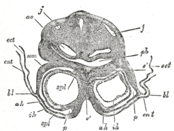 Transverse section through the region of the heart in a rabbit embryo of nine days. X 80. (Kölliker.) j, j. Jugular veins. ao. Aorta. ph. Pharynx. som. Somatopleure. bl. Proamnion. ect. Ectoderm. ent. Entoderm. p. Pericardium. spl. Splanchnopleure. ah. Outer wall of heart. ih. Endothelial lining of heart. é. Septum between heart tubes. | |
| Details | |
| Carnegie stage | 9 |
| Precursor | lateral plate mesoderm, ectoderm |
| Gives rise to | mesenchyme |
| Identifiers | |
| Latin | mesenchyma somatopleurale |
| TE | E4.0.4.1.0.0.3 |
| Anatomical terminology | |
See also
References
This article incorporates text in the public domain from page 50 of the 20th edition of Gray's Anatomy (1918)
External links
- Diagram at Yuba Community College District at the Library of Congress Web Archives (archived 2008-08-09)
- Overview at Kennesaw State University
| Authority control |
|
|---|
This article is issued from
Wikipedia.
The text is licensed under Creative
Commons - Attribution - Sharealike.
Additional terms may apply for the media files.