Solid pseudopapillary tumour
A solid pseudopapillary tumour is a low-grade malignant neoplasm of the pancreas of papillary architecture that typically afflicts young women.[1]
| Solid pseudopapillary tumour | |
|---|---|
| Other names | Solid pseudopapillary neoplasm, solid pseudopapillary tumour/neoplasm of the pancreas, Frantz's tumour |
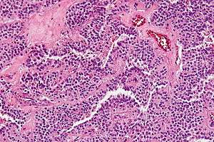 | |
| Micrograph of a solid pseudopapillary tumour. H&E stain. | |
| Specialty | Oncology |
Gross morphology
Solid pseudopapillary tumours are typically round, well-demarcated, measuring 2–17 cm in diameter (average 8 cm), with solid and cystic areas with hemorrhage on cut sections.[2]
Histomorphology
Solid pseudopapillary tumours consist of solid sheets of cells that are focally dyscohesive. The cells in the lesion usually have uniform nuclei with occasional nuclear grooves, eosinophilic or clear cytoplasm and PAS positive eosinophilic intracytoplasmic globules.[3] Necrosis is usually present and, as cell death preferentially occurs distant from blood vessels, lead to the formation of pseudopapillae.
Immunohistochemistry
Solid pseudopapillary tumours show positive nuclear staining for beta catenin, as well as positive immunostaining for CD10, CD56, vimentin, alpha 1-antitrypsin, and neuron specific enolase; they are negative for chromogranin and pancreatic enzymes.[4][5]
See also
- Pancreatic cancer
- Pancreatic mucinous cystic neoplasm
- Serous cystadenoma of the pancreas
References
- Patil TB, Shrikhande SV, Kanhere HA, Saoji RR, Ramadwar MR, Shukla PJ (2006). "Solid pseudopapillary neoplasm of the pancreas: a single institution experience of 14 cases". HPB. 8 (2): 148–50. doi:10.1080/13651820510035721. PMC 2131425. PMID 18333264.
- Fletcher CDM (2007). Diagnostic Histopathology of Tumors. I (3rd ed.). Elsevier. p. 478.
- Serra S, Chetty R (November 2008). "Revision 2: an immunohistochemical approach and evaluation of solid pseudopapillary tumour of the pancreas". J. Clin. Pathol. 61 (11): 1153–9. doi:10.1136/jcp.2008.057828. PMID 18708424.
- Stömmer P, Kraus J, Stolte M, Giedl J (March 1991). "Solid and cystic pancreatic tumors. Clinical, histochemical, and electron microscopic features in ten cases". Cancer. 67 (6): 1635–41. doi:10.1002/1097-0142(19910315)67:6<1635::aid-cncr2820670627>3.0.co;2-m. PMID 1900454.
- Pettinato G, Manivel JC, Ravetto C, et al. (November 1992). "Papillary cystic tumor of the pancreas. A clinicopathologic study of 20 cases with cytologic, immunohistochemical, ultrastructural, and flow cytometric observations, and a review of the literature". Am. J. Clin. Pathol. 98 (5): 478–88. PMID 1283055.
External links
| Classification |
|---|