Cervical sinus
During Human embryogenesis the mandibular arch and hyoid arch grow more rapidly than those behind them, with the result that the latter become, to a certain extent, telescoped within the former, and a deep depression, the cervical sinus, is formed on either side of the neck.
| Cervical sinus | |
|---|---|
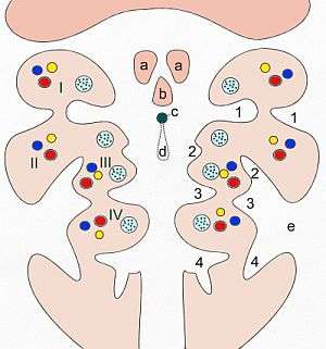 Pattern of the branchial arches. I-IV branchial arches, 1-4 branchial pouches (inside) and/or pharyngeal grooves (outside) a Tuberculum laterale b Tuberculum impar c Foramen cecum d Ductus thyreoglossus e Sinus cervicalis | |
| Details | |
| Identifiers | |
| Latin | sinus cervicalis |
| Anatomical terminology | |
This sinus is bounded in front by the hyoid arch, and behind by the thoracic wall; it is ultimately obliterated by the fusion of its walls.
Formed by the walls of the 2-4th pharyngeal grooves merging. Sometimes it can remain anterior to the sternocleidomastoid muscle. It can communicate with the skin( external cervical fistula) or with the pharynx (internal cervical fistula). Prone to infection.
Additional images
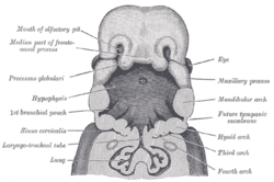 The head and neck of a human embryo thirty-two days old, seen from the ventral surface.
References
This article incorporates text in the public domain from page 67 of the 20th edition of Gray's Anatomy (1918)