Radial keratotomy
Radial keratotomy (RK) is a refractive surgical procedure to correct myopia (nearsightedness) that was developed in 1974, by Svyatoslav Fyodorov, a Russian ophthalmologist. It has been largely supplanted by newer operations, such as photorefractive keratectomy, LASIK, Epi-LASIK and the phakic intraocular lens.
| Radial keratotomy | |
|---|---|
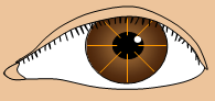 Schematic diagram of RK, with incisions drawn in orange | |
| ICD-9-CM | 11 |
| MeSH | D007646 |
History
Beginning in 1936, Japanese ophthalmologist Tsutomu Sato conducted research in anterior and posterior keratotomy, an early form of refractive surgery that attempted to treat keratoconus, myopia and astigmatism by making incisions in the cornea.[1]
In 1974, Svyatoslav Fyodorov removed glass from the eye of a boy who had been in an accident. The boy, who required eyeglasses for correction of myopia caused by astigmatism, fell off his bicycle. His glasses shattered on impact, and glass particles lodged in both eyes. To save the boy's vision, Fyodorov performed an operation which consisted of making numerous radial incisions extending from the pupil to the periphery of the cornea in a radial pattern like the spokes of a wheel. After the glass was removed by this method and the cornea healed, Fyodorov found that the boy's visual acuity had improved significantly.
Procedure
In RK, incisions are made with a diamond knife. Incisions that penetrate only the superficial corneal stroma are less effective than those reaching deep into the cornea,[2] and consequently, incisions are made quite deep. One study cites incisions made to a depth equivalent to the thinnest of four corneal-thickness measurements made near the center of the cornea.[3] Other sources cite surgeries leaving 20 to 50 micrometres of corneal tissue unincised (roughly equivalent to 90% of corneal depth, based on thickness norms).[2]
Results
Visual acuity is generally improved.[4]
Postsurgical healing
The healing corneal wounds consist of newly abutting corneal stroma, fibroblastic cells, and irregular fibrous connective tissue. Closer to the wound surface lies the epithelial plug, a bed of the cells that form the normal corneal epithelium which have fallen into the wound. Often this plug is three to four times as deep as the normal corneal epithelium layer. As the cells migrate from the depth of the plug up to the surface, some die before reaching it, forming breaches in the otherwise healthy epithelial layer. This, consequently, leaves the cornea more susceptible to infections.[5][6][7] The risk is estimated to be between 0.25%[8] and 0.7%[9] Healing of the RK incisions is very slow and unpredictable, often incomplete even years after surgery.[10] Similarly, infection of these chronic wounds can also occur years after surgery,[11][12][13] with 53% of ocular infections being late in onset.[14] The pathogen most commonly involved in such infections is the highly virulent bacterium Pseudomonas aeruginosa.[15]
Complications
Large epithelial plugs may cause more scattering of light, leading to the appearance of visual phenomena such as flares and starbursts — especially in situations such as night driving, where the stark light of car headlights abounds. These dark conditions cause the pupil to dilate, maximizing the amount of scattered light that enters the eye. In cases where large epithelial plugs lead to such aggravating symptoms, patients may seek further surgical treatment to alleviate the symptoms.[5]
Increasing altitude can cause partial blindness in people who have undergone RK, as discovered by mountaineer Beck Weathers (who had undergone RK) during the 1996 Mount Everest disaster.
The incisions of RK are used to relax the steep central cornea in patients with myopia. The original technique — consisting of incisions from periphery to center — was called "the Russian technique",[16] while the later advances of performing controlled incision from center to periphery was called "the American technique".[17]
RK enjoyed great popularity during the 1980s, and was one of the most studied refractive surgical procedures. Its 10-year data was published as the PERK (Prospective Evaluation of Radial Keratotomy) study, which proved the onset of progressive hyperopia — often found a decade after the original surgery — is due to continued flattening of the central cornea.
A conceptually opposite technique of using hexagonal incisions in the periphery of the cornea is known has hexagonal keratotomy (HK, described by Antonio Mendez of Mexicali, Mexico), which was used to correct low degrees of hyperopia. The idea behind HK was to make six peripheral incisions forming a hexagon around the central cornea to steepen the hyperopic flat cornea and, thereby, focus the rays of light more precisely onto the retina. These incisions can be of two types, either connecting or non-connecting.[18]
RK may be performed with different types, numbers, and patterns of incisions. They can have 4, 8, 12, 16 or 32 incisions made in a number of patterns and orientations based on refractive errors, surgeon style and surgeon training. Many of these patients have had additional incisional surgeries like astigmatic keratotomy (AK), where incisions are placed at the steepest points of the cornea in people with astigmatism to relax and transform the cornea to a more spherical shape. Some people have had a combination of intraocular surgeries, such as pseudophakic or phakic implants, along with their keratotomies and many underwent "purse-string" suturing to control over-correction (Dr. Green’s lasso suture).
Due to the instability of the cornea seen with many age-related pathologies, it may be difficult to address visual acuity satisfactorily in people who have undergone RK surgery but who later develop presbyopia (hyperopia caused by age-related changes in the crystalline lens). In these situations, factors to be considered include:
Primary visual factors:
- Quantitative:
- Decreased visual acuity (myopia, hyperopia, astigmatism)
- Qualitative:
- Irregular astigmatism
- Small optic zone
- Incisions
Secondary (associated) visual factors:
- Presbyopia
- Cataracts
- Corneal scars
- Corneal instability (thin/ectasia/trampoline effect)
Visual rehabilitation after RK
The PERK study demonstrated that people who undergo RK continue to drift toward hyperopia ("farsightedness"). Additionally, many of these people have reached the age where presbyopia occurs. Some also develop cataracts. Their vision can still be restored with Epi-LASIK, photorefractive keratectomy, LASIK or phakic lens extraction, or cataract surgery. The corneal curvature has to remeasured and modified by history, central keratometry, or contact lens method.
References
- Sato, T (1939). "Treatment of conical cornea (incision of Descemet's membrane)". Acta Soc Ophthalmol Jpn (in Japanese). 43: 544–55.
- Bashour M, Benchimol M. (2005) Emedicine. Viewed 12 October 2006. Myopia, Radial Keratotomy
- Waring GO, Moffitt SD, Gelender H, et al. (January 1983). "Rationale for and design of the National Eye Institute Prospective Evaluation of Radial Keratotomy (PERK) Study". Ophthalmology. 90 (1): 40–58. doi:10.1016/s0161-6420(83)34603-0. PMID 6338438.
- Symons SP, Slomovic AR (August 1994). "Visual acuity, refractive and keratometric results of 140 consecutive radial keratotomy procedures". Canadian Journal of Ophthalmology. 29 (4): 176–181.
- Bergmanson JP, Farmer EJ (1999). "A return to primitive practice? Radial keratotomy revisited". Cont Lens Anterior Eye. 22 (1): 2–10. doi:10.1016/S1367-0484(99)80024-1. PMID 16303397.
- Bergmanson J, Farmer E, Goosey J (November 2001). "Epithelial plugs in radial keratotomy: the origin of incisional keratitis?". Cornea. 20 (8): 866–72. doi:10.1097/00003226-200111000-00018. PMID 11685068.
- Deg JK, Zavala EY, Binder PS (June 1985). "Delayed corneal wound healing following radial keratotomy". Ophthalmology. 92 (6): 734–40. doi:10.1016/s0161-6420(85)33963-5. PMID 4034168.
- Waring GO, Lynn MJ, McDonnell PJ (October 1994). "Results of the prospective evaluation of radial keratotomy (PERK) study 10 years after surgery". Arch. Ophthalmol. 112 (10): 1298–308. doi:10.1001/archopht.1994.01090220048022. PMID 7945032.
- Hoffer KJ, Darin JJ, Pettit TH, Hofbauer JD, Elander R, Levenson JE (June 1983). "Three years experience with radial keratotomy. The UCLA study". Ophthalmology. 90 (6): 627–36. doi:10.1016/s0161-6420(83)34517-6. PMID 6350968.
- Binder PS, Nayak SK, Deg JK, Zavala EY, Sugar J (March 1987). "An ultrastructural and histochemical study of long-term wound healing after radial keratotomy". Am. J. Ophthalmol. 103 (3 Pt 2): 432–40. doi:10.1016/s0002-9394(14)77767-0. PMID 3826260.
- McClellan KA, Bernard PJ, Gregory-Roberts JC, Billson FA (May 1988). "Suppurative keratitis: a late complication of radial keratotomy". J Cataract Refract Surg. 14 (3): 317–20. doi:10.1016/s0886-3350(88)80124-x. PMID 3397895.
- Mandelbaum S, Waring GO, Forster RK, Culbertson WW, Rowsey JJ, Espinal ME (August 1986). "Late development of ulcerative keratitis in radial keratotomy scars". Arch. Ophthalmol. 104 (8): 1156–60. doi:10.1001/archopht.1986.01050200062050. PMID 3741245.
- Wilhelmus K, Hanburg S (June 1983). "Bacterial Keratitis following Radial Keratotomy". Cornea. 2 (2): 143–6. doi:10.1097/00003226-198302020-00009.
- Jain S, Azar DT (1996). "Eye infections after refractive keratotomy". J Refract Surg. 12 (1): 148–55. PMID 8963804.
- Heidemann DG, Dunn SP, Chow CY (December 1999). "Early- versus late-onset infectious keratitis after radial and astigmatic keratotomy: clinical spectrum in a referral practice". J Cataract Refract Surg. 25 (12): 1615–9. doi:10.1016/S0886-3350(99)00285-0. PMID 10609205.
- Gulani AC, Fyodorov S: Future Directions in Vision course, June 1997
- Gulani AC, Neumann AC: Refractive Surgery Course, February 1996
- Gulani AC: 10 Refractive Procedures for Hyperopia. ISOPT 2001