Nonunion
Nonunion is permanent failure of healing following a broken bone unless intervention (such as surgery) is performed. A fracture with nonunion generally forms a structural resemblance to a fibrous joint, and is therefore often called a "false joint" or pseudoarthrosis (the Greek stem "pseudo-" means false and "arthrosis" means joint). The diagnosis is generally made when there is no healing between two sets of medical imaging such as X-ray or CT scan. This is generally after 6–8 months.[1]
| Nonunion | |
|---|---|
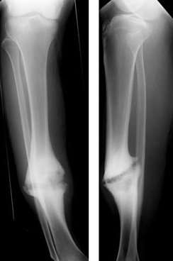 | |
| Hypertrophic nonunion of the tibia | |
| Specialty | Orthopedics |
Nonunion is a serious complication of a fracture and may occur when the fracture moves too much, has a poor blood supply or gets infected. Patients who smoke have a higher incidence of nonunion. The normal process of bone healing is interrupted or stalled.
Since the process of bone healing is quite variable, a nonunion may go on to heal without intervention in a very few cases. In general, if a nonunion is still evident at 6 months post injury it will remain unhealed without specific treatment, usually orthopedic surgery. A non-union which does go on to heal is called a delayed union.[2]
Signs and symptoms
A history of a broken bone is usually apparent. The patient complains of persistent pain at the fracture site and may also notice abnormal movement or clicking at the level of the fracture. An x-ray plate of the fractured bone shows a persistent radiolucent line at the fracture. Callus formation may be evident but callus does not bridge across the fracture. If there is doubt about the interpretation of the x-ray, stress x-rays, tomograms or CT scan may be used for confirmation.
Cause
The reasons for non-union are
- avascular necrosis (the blood supply was interrupted by the fracture)
- the two ends are not apposed (that is, they are not next to each other)
- infection (particularly osteomyelitis)
- the fracture is not fixed (that is, the two ends are still mobile)
- soft-tissue imposition (there is muscle or ligament covering the broken ends and preventing them from touching each other)
Risk factors
- Related to the person:
- Age: Common in old age
- Nutritional status : poor
- Habits : Nicotine and alcohol consumption
- Metabolic disturbance : Hyperparathyroidism
- can be found in those with NF1
- Genetic predisposition[3]
- Causes related to fracture:
- Related to the fracture site
- Soft tissue interposition
- Bone loss at the fracture
- Infection
- Loss of blood supply
- Damage of surrounding muscles
- Related to treatment
- Inadequate reduction
- Insufficient immobilization
- Improperly applied fixation devices.
Hypertrophic non-union
Callus is formed, but the bone fractures have not joined. This can be due to inadequate fixation of the fracture, and treated with rigid immobilisation.
Atrophic non-union
No callus is formed. This is often due to impaired bony healing, for example due to vascular causes (e.g. impaired blood supply to the bone fragments) or metabolic causes (e.g. diabetes or smoking). Failure of initial union, for example when bone fragments are separated by soft tissue may also lead to atrophic non-union. Atrophic non-union can be treated by improving fixation, removing the end layer of bone to provide raw ends for healing, and the use of bone grafts.
Diagnosis
The diagnosis of nonunion is generally done when there is no progress between two occasions of medical imaging such as X-ray. This is generally the case after 6–8 months.[1]
Types of Nonunion
Judet and Judet, Muller, Weber and Cech, and others classified nonunions into two types according to the viability of the ends of the fragments: Hypervascular nonunions and avascular nonunions.
Hypervascular nonunions are subdivided as:
- "Elephant foot" nonunions: These are hypertrophic, rich in callus and are a result of inadequate immobilisation, insecure fixation or premature weight bearing.
- "Horse hoof" nonunions: Mildly hypertrophic, poor in callus and is due to unstable fixation.
- Oligotrophic nonunions: They are not hypertrophic but vascular, no callus seen and is due to severely displaced fracture or fixation without accurate apposition of fragments.
Avascular nonunions are subdivided as:
- Torsion wedge nonunions have an intermediate fragment with decreased or absent blood supply. This fragment has healed to one main fragment but not to the other.
- Comminuted nonunions have one or more intermediate fragments that are necrotic.
- Defect nonunions has a gap in diaphysis of bone due to a loss of fragment.
- Atrophic nonunions usually are the final result when the intermediate fragments are missing and scar tissue that lacks osteogenic potential is left in their place.
Paley classified tibial nonunions based on clinical and roentgenographic characteristics as Type A (Bone loss of less than 1 cm) and Type B (Bone loss of more than 1 cm). Type A is subclassified as Type A:1 Lax type; Lax nonunion have limited mobility and usually some fixed deformity, Type A:2:1 stiff nonunion without deformity and Type A:2:2 stiff nonunion with a deformity. Type B subclassified as Type B:1 bony defect with no shortening, Type B:2 shortening with no gap and Type B:3 there is both gap and shortening.
Treatment
Surgery
Surgical treatment options include:
- Removal of all scar tissue from between the fracture fragments
- Immobilization of the fracture with internal or external fixation. Metal plates, pins, screws, and rods, that are screwed or driven into a bone, are used to stabilize the broken bone fragments.
- Bone grafting. Donor bone or autologous bone (harvested from the same person undergoing the surgery) is used as a stimulus to bone healing. The presence of the bone is thought to cause stem cells in the circulation and marrow to form cartilage, which then turns to bone, instead of a fibrous scar that forms to heal all other tissues of the body. Bone is the only tissue that can heal without a fibrous scar. Autologus bone graft is the "gold standard" treatment of the non union the bone is obtained from the iliac crest.
In simple cases healing may be evident within 3 months. Gavriil Ilizarov revolutionized the treatment of recalcitrant nonunions demonstrating that the affected area of the bone could be removed, the fresh ends "docked" and the remaining bone lengthened using an external fixator device.[4] The time course of healing after such treatment is longer than normal bone healing. Usually there are signs of union within 3 months, but the treatment may continue for many months beyond that.
Bone stimulation
Bone stimulation may be with either electromagnetic or ultrasound waves.[5] Ultrasound stimulation has tentative evidence of supporting better healing in long bones that have not healed after three months.[6] Evidence; from a Cochrane review however, does not show that ultrasound decreases rates of nonunion.[7] Another review has, however, suggested it as an alternative to surgery.[8]
Prognosis
By definition, a nonunion will not heal if left alone. Therefore the patient's symptoms will not be improved and the function of the limb will remain impaired. It will be painful to bear weight on it and it may be deformed or unstable. The prognosis of nonunion if treated depends on many factors including the age and general health of the patient, the time since the original injury, the number of previous surgeries, smoking history, the patient's ability to cooperate with the treatment. In the region of 80% of nonunions heal after the first operation. The success rate with subsequent surgeries is less.
See also
References
- Page 542 in: Rigmor Texhammar, Christopher Colton (2013). AO/ASIF Instruments and Implants: A Technical Manual (2 ed.). Springer Science & Business Media. ISBN 9783662030325.
- "Nonunions - OrthoInfo - AAOS". Retrieved 2018-09-02.
- McCoy, Thomas H.; Fragomen, Austin T.; Hart, Kamber L.; Pellegrini, Amelia M.; Raskin, Kevin A.; Perlis, Roy H. (January 2019). "Genomewide Association Study of Fracture Nonunion Using Electronic Health Records". JBMR Plus. 3 (1): 23–28. doi:10.1002/jbm4.10063. ISSN 2473-4039. PMC 6339539. PMID 30680360.
- Niedzielski K, Synder M (2000). "The treatment of pseudarthrosis using the Ilizarov method". Ortop Traumatol Rehabil. 2 (3): 46–8. PMID 18034140.
- Victoria, Galkowski; Petrisor, Brad; Drew, Brian; Dick, David (2009). "Bone stimulation for fracture healing: What′s all the fuss?". Indian Journal of Orthopaedics. 43 (2): 117–20. doi:10.4103/0019-5413.50844. ISSN 0019-5413. PMC 2762251. PMID 19838359.
- Higgins, A; Glover, M; Yang, Y; Bayliss, S; Meads, C; Lord, J (October 2014). "EXOGEN ultrasound bone healing system for long bone fractures with non-union or delayed healing: a NICE medical technology guidance". Applied Health Economics and Health Policy. 12 (5): 477–84. doi:10.1007/s40258-014-0117-6. PMC 4175405. PMID 25060830.
- Griffin, XL; Parsons, N; Costa, ML; Metcalfe, D (23 June 2014). "Ultrasound and shockwave therapy for acute fractures in adults". The Cochrane Database of Systematic Reviews (6): CD008579. doi:10.1002/14651858.CD008579.pub3. PMID 24956457.
- Leighton, R.; Watson, J.T; Giannoudis, P.; Papakostidis, C.; Harrison, A.; Steen, R.G. (May 2017). "Healing of fracture nonunions treated with low-intensity pulsed ultrasound (LIPUS): A systematic review and meta-analysis" (PDF). Injury. 48 (7): 1339–1347. doi:10.1016/j.injury.2017.05.016. PMID 28532896.