Protozoa
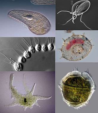
Protozoa (also protozoan, plural protozoans) is an informal term for single-celled eukaryotes, either free-living or parasitic, which feed on organic matter such as other microorganisms or organic tissues and debris.[1][2] Historically, the protozoa were regarded as "one-celled animals", because they often possess animal-like behaviors, such as motility and predation, and lack a cell wall, as found in plants and many algae.[3][4] Although the traditional practice of grouping protozoa with animals is no longer considered valid, the term continues to be used in a loose way to identify single-celled organisms that can move independently and feed by heterotrophy.
In some systems of biological classification, Protozoa is a high-level taxonomic group. When first introduced in 1818, Protozoa was erected as a taxonomic class,[5] but in later classification schemes it was elevated to a variety of higher ranks, including phylum, subkingdom and kingdom. In a series of classifications proposed by Thomas Cavalier-Smith and his collaborators since 1981, Protozoa has been ranked as a kingdom.[6][7][8] The seven-kingdom scheme presented by Ruggiero et al. in 2015, places eight phyla under Kingdom Protozoa: Euglenozoa, Amoebozoa, Metamonada, Choanozoa sensu Cavalier-Smith, Loukozoa, Percolozoa, Microsporidia and Sulcozoa.[9] Notably, this kingdom excludes several major groups of organisms traditionally placed among the protozoa, including the ciliates, dinoflagellates, foraminifera, and the parasitic apicomplexans, all of which are classified under Kingdom Chromista. Kingdom Protozoa, as defined in this scheme, does not form a natural group or clade, but a paraphyletic group or evolutionary grade, within which the members of Fungi, Animalia and Chromista are thought to have evolved.[9]
History
The word "protozoa" (singular protozoon or protozoan) was coined in 1818 by zoologist Georg August Goldfuss, as the Greek equivalent of the German Urthiere, meaning "primitive, or original animals" (ur- ‘proto-’ + Thier ‘animal’). Goldfuss created Protozoa as a class containing what he believed to be the simplest animals.[5] Originally, the group included not only single-celled microorganisms but also some "lower" multicellular animals, such as rotifers, corals, sponges, jellyfish, bryozoa and polychaete worms.[10] The term Protozoa is formed from the Greek words πρῶτος (prôtos), meaning "first", and ζῶα (zôa), plural of ζῶον (zôon), meaning "animal".[11][12] The use of Protozoa as a formal taxon has been discouraged by some researchers, mainly because the term implies kinship with animals (Metazoa)[13][14] and promotes an arbitrary separation of "animal-like" from "plant-like" organisms.[15]
In 1848, as a result of advancements in cell theory pioneered by Theodor Schwann and Matthias Schleiden, the anatomist and zoologist C. T. von Siebold proposed that the bodies of protozoans such as ciliates and amoebae consisted of single cells, similar to those from which the multicellular tissues of plants and animals were constructed. Von Siebold redefined Protozoa to include only such unicellular forms, to the exclusion of all metazoa (animals).[16] At the same time, he raised the group to the level of a phylum containing two broad classes of microorganisms: Infusoria (mostly ciliates and flagellated algae), and Rhizopoda (amoeboid organisms). The definition of Protozoa as a phylum or sub-kingdom composed of "unicellular animals" was adopted by the zoologist Otto Bütschli—celebrated at his centenary as the "architect of protozoology"[17]—and the term came into wide use.
As a phylum under Animalia, the Protozoa were firmly rooted in the old "two-kingdom" classification of life, according to which all living beings were classified as either animals or plants. As long as this scheme remained dominant, the protozoa were understood to be animals and studied in departments of Zoology, while photosynthetic microorganisms and microscopic fungi—the so-called Protophyta—were assigned to the Plants, and studied in departments of Botany.[18]
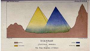
Criticism of this system began in the latter half of the 19th century, with the realization that many organisms met the criteria for inclusion among both plants and animals. For example, the algae Euglena and Dinobryon have chloroplasts for photosynthesis, but can also feed on organic matter and are motile. In 1860, John Hogg argued against the use of "protozoa", on the grounds that "naturalists are divided in opinion—and probably some will ever continue so—whether many of these organisms, or living beings, are animals or plants."[13] As an alternative, he proposed a new kingdom called Primigenum, consisting of both the protozoa and unicellular algae (protophyta), which he combined together under the name "Protoctista". In Hoggs's conception, the animal and plant kingdoms were likened to two great "pyramids" blending at their bases in the Kingdom Primigenum.
Six years later, Ernst Haeckel also proposed a third kingdom of life, which he named Protista. At first, Haeckel included a few multicellular organisms in this kingdom, but in later work he restricted the Protista to single-celled organisms, or simple colonies whose individual cells are not differentiated into different kinds of tissues.
Despite these proposals, Protozoa emerged as the preferred taxonomic placement for heterotrophic microorganisms such as amoebae and ciliates, and remained so for more than a century. In the course of the 20th century, however, the old "two kingdom" system began to weaken, with the growing awareness that fungi did not belong among the plants, and that most of the unicellular protozoa were no more closely related to the animals than they were to the plants. By mid-century, some biologists, such as Herbert Copeland, Robert H. Whittaker and Lynn Margulis, advocated the revival of Haeckel's Protista or Hogg's Protoctista as a kingdom-level eukaryotic group, alongside Plants, Animals and Fungi.[18] A variety of multi-kingdom systems were proposed, and Kingdoms Protista and Protoctista became well established in biology texts and curricula.[19][20][21]
While many taxonomists have abandoned Protozoa as a high-level group, Thomas Cavalier-Smith has retained it as a kingdom in the various classifications he has proposed. As of 2015, Cavalier-Smith's Protozoa excludes several major groups of organisms traditionally placed among the protozoa, including the ciliates, dinoflagellates and foraminifera (all members of the SAR supergroup). In its current form, his kingdom Protozoa is a paraphyletic group which includes a common ancestor and most of its descendants, but excludes two important clades that branch within it: the animals and fungi.[9]
Since the protozoa, as traditionally defined, can no longer be regarded as "primitive animals" the terms "protists", "Protista" or "Protoctista" are sometimes preferred. In 2005, members of the Society of Protozoologists voted to change its name to the International Society of Protistologists.[22]
Characteristics
Size
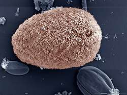
Protozoa, as traditionally defined, range in size from as little as 1 micrometre to several millimetres, or more.[23] Among the largest are the deep-sea–dwelling xenophyophores, single-celled foraminifera whose shells can reach 20 cm in diameter.[24]
| Species | Cell type | Size in micrometres |
|---|---|---|
| Plasmodium falciparum | malaria parasite, trophozoite phase[25] | 1-2 |
| Massisteria voersi | free-living cercozoan amoeboid[26] | 2.3–3 |
| Bodo saltans | free living kinetoplastid flagellate[27] | 5-8 |
| Plasmodium falciparum | malaria parasite, gametocyte phase[28] | 7-14 |
| Trypanosoma cruzi | parasitic kinetoplastid, Chagas disease[29] | 14-24 |
| Entamoeba histolytica | parasitic amoebozoan[30] | 15–60 |
| Balantidium coli | parasitic ciliate[31] | 50-100 |
| Paramecium caudatum | free-living ciliate[32] | 120-330 |
| Amoeba proteus | free-living amoebozoan[33] | 220–760 |
| Noctiluca scintillans | free-living dinoflagellate[34] | 700–2000 |
| Syringammina fragilissima | foraminiferan amoeboid[24] | up to 200000 |
Habitat
Free-living protozoans are common and often abundant in fresh, brackish and salt water, as well as other moist environments, such as soils and mosses. Some species thrive in extreme environments such as hot springs[35] and hypersaline lakes and lagoons.[36] All protozoa require a moist habitat; however, some can survive for long periods of time in dry environments, by forming resting cysts which enable them to remain dormant until conditions improve.
Parasitic and symbiotic protozoa live on or within other organisms, including vertebrates and invertebrates, as well as plants and other single-celled organisms. Some are harmless or beneficial to their host organisms; others may be significant causes of diseases, such as babesia, malaria and toxoplasmosis.
Association between protozoan symbionts and their host organisms can be mutually beneficial. Flagellated protozoans such as Trichonympha and Pyrsonympha inhabit the guts of termites, where they enable their insect host to digest wood by helping to break down complex sugars into smaller, more easily digested molecules.[37] A wide range of protozoans live commensally in the rumens of ruminant animals, such as cattle and sheep. These include flagellates, such as Trichomonas, and ciliated protozoa, such as Isotricha and Entodinium.[38] The ciliate subclass Astomatia is composed entirely of mouthless symbionts adapted for life in the guts of annelid worms.[39]
Feeding
All protozoans are heterotrophic, deriving nutrients from other organisms, either by ingesting them whole or consuming their organic remains and waste-products. Some protozoans take in food by phagocytosis, engulfing organic particles with pseudopodia (as amoebae do), or taking in food through a specialized mouth-like aperture called a cytostome. Others take in food by osmotrophy, absorbing dissolved nutrients through their cell membranes.
Parasitic protozoans use a wide variety of feeding strategies, and some may change methods of feeding in different phases of their life cycle. For instance, the malaria parasite Plasmodium feeds by pinocytosis during its immature trophozoite stage of life (ring phase), but develops a dedicated feeding organelle (cytostome) as it matures within a host's red blood cell.[40]

Protozoa may also live as mixotrophs, supplementing a heterotrophic diet with some form of autotrophy. Some protozoa form close associations with symbiotic photosynthetic algae, which live and grow within the membranes of the larger cell and provide nutrients to the host. Others practice kleptoplasty, stealing chloroplasts from prey organisms and maintaining them within their own cell bodies as they continue to produce nutrients through photosynthesis. The ciliate Mesodinium rubrum retains functioning plastids from the cryptophyte algae on which it feeds, using them to nourish themselves by autotrophy. These, in turn, may be passed along to dinoflagellates of the genus Dinophysis , which prey on Mesodinium rubrum but keep the enslaved plastids for themselves. Within Dinophysis, these plastids can continue to function for months.[41]
Motility
Organisms traditionally classified as protozoa are abundant in aqueous environments and soil, occupying a range of trophic levels. The group includes flagellates (which move with the help of whip-like structures called flagella), ciliates (which move by using hair-like structures called cilia) and amoebae (which move by the use of foot-like structures called pseudopodia). Some protozoa are sessile, and do not move at all.
Pellicle
Unlike plants, fungi and most types of algae, protozoans do not typically have a rigid cell wall, but are usually enveloped by elastic structures of membranes that permit movement of the cell. In some protozoans, such as the ciliates and euglenozoans, the cell is supported by a composite membranous envelope called the "pellicle". The pellicle gives some shape to the cell, especially during locomotion. Pellicles of protozoan organisms vary from flexible and elastic to fairly rigid. In ciliates and Apicomplexa, the pellicle is supported by closely packed vesicles called alveoli. In euglenids, it is formed from protein strips arranged spirally along the length of the body. Familiar examples of protists with a pellicle are the euglenoids and the ciliate Paramecium. In some protozoa, the pellicle hosts epibiotic bacteria that adhere to the surface by their fimbriae (attachment pili).[42]
Life cycle
Some protozoa have two-phase life cycles, alternating between proliferative stages (e.g., trophozoites) and dormant cysts. As cysts, protozoa can survive harsh conditions, such as exposure to extreme temperatures or harmful chemicals, or long periods without access to nutrients, water, or oxygen for periods of time. Being a cyst enables parasitic species to survive outside of a host, and allows their transmission from one host to another. When protozoa are in the form of trophozoites (Greek tropho = to nourish), they actively feed. The conversion of a trophozoite to cyst form is known as encystation, while the process of transforming back into a trophozoite is known as excystation.
All protozoans reproduce (not all) asexually by binary fission or multiple fission. Many protozoan species exchange genetic material by sexual means (typically, through conjugation); however, sexuality is generally decoupled from the process of reproduction, and does not immediately result in increased population.[43]
Although meiotic sex is widespread among present day eukaryotes, it has, until recently, been unclear whether or not eukaryotes were sexual early in their evolution. Due to recent advances in gene detection and other techniques, evidence has been found for some form of meiotic sex in an increasing number of protozoans of ancient lineage that diverged early in eukaryotic evolution.[44] (See eukaryote reproduction.) Thus, such findings suggest that meiotic sex arose early in eukaryotic evolution. Examples of protozoan meiotic sexuality are described in the articles Amoebozoa, Giardia lamblia, Leishmania, Plasmodium falciparum biology, Paramecium, Toxoplasma gondii, Trichomonas vaginalis and Trypanosoma brucei.
Classification
Historically, the Protozoa were classified as "unicellular animals", as distinct from the Protophyta, single-celled photosynthetic organisms (algae) which were considered primitive plants. Both groups were commonly given the rank of phylum, under the kingdom Protista.[45] In older systems of classification, the phylum Protozoa was commonly divided into several sub-groups, reflecting the means of locomotion.[46] Classification schemes differed, but throughout much of the 20th century the major groups of Protozoa included:
- Flagellates, or Mastigophora (motile cells equipped with whiplike organelles of locomotion, e.g., Giardia lamblia)
- Amoebae or Sarcodina (cells that move by extending pseudopodia or lamellipodia, e.g., Entamoeba histolytica)
- Sporozoans, or Sporozoa (parasitic, spore-producing cells, whose adult form lacks organs of motility, e.g., Plasmodium knowlesi)
- Apicomplexa (now in Alveolata)
- Microsporidia (now in Fungi)
- Ascetosporea (now in Rhizaria)
- Myxosporidia (now in Cnidaria)
- Ciliates, or Ciliophora (cells equipped with large numbers of short hairlike organs of locomotion, e.g., Balantidium coli
With the emergence of molecular phylogenetics and tools enabling researchers to directly compare the DNA of different organisms, it became evident that, of the main sub-groups of Protozoa, only the ciliates (Ciliophora) formed a natural group, or monophyletic clade (that is, a distinct lineage of organisms sharing common ancestry). The other classes or subphyla of Protozoa were all polyphyletic groups composed of organisms that, despite similarities of appearance or way of life, were not necessarily closely related to one another. In the system of eukaryote classification currently endorsed by the International Society of Protistologists, members of the old phylum Protozoa have been distributed among a variety of supergroups.[47]
Ecology
As components of the micro- and meiofauna, protozoa are an important food source for microinvertebrates. Thus, the ecological role of protozoa in the transfer of bacterial and algal production to successive trophic levels is important. As predators, they prey upon unicellular or filamentous algae, bacteria, and microfungi. Protozoan species include both herbivores and consumers in the decomposer link of the food chain. They also control bacteria populations and biomass to some extent.
Disease
A number of protozoan pathogens are human parasites, causing diseases such as malaria (by Plasmodium), amoebiasis, giardiasis, toxoplasmosis, cryptosporidiosis, trichomoniasis, Chagas disease, leishmaniasis, African trypanosomiasis (sleeping sickness), amoebic dysentery, acanthamoeba keratitis, and primary amoebic meningoencephalitis (naegleriasis).
The protozoan Ophryocystis elektroscirrha is a parasite of butterfly larvae, passed from female to caterpillar. Severely infected individuals are weak, unable to expand their wings, or unable to eclose, and have shortened lifespans, but parasite levels vary in populations. Infection creates a culling effect, whereby infected migrating animals are less likely to complete the migration. This results in populations with lower parasite loads at the end of the migration.[48] This is not the case in laboratory or commercial rearing, where after a few generations, all individuals can be infected.[49]
List of protozoan diseases in humans:[50]
| Disease | Causative agent | Source of Transmission |
|---|---|---|
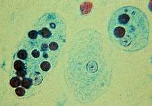
| Amoebiasis | Entamoeba histolytica (Amoebozoa) | Water, food |
| Acanthamoeba keratitis | Acanthamoeba (Amoebozoa) | Water, contaminated contact lens solution |
| Giardiasis | Giardia lamblia (Metamonada) | Water, Contact |
| Trichomoniasis | Trichomonas vaginalis (Metamonada) | Sexual contact |
| Dientamoebiasis | Dientamoeba fragilis (Metamonada) | Uncertain |
| African sleeping sickness (African trypanosomiasis) | Trypanosoma brucei (Kinetoplastida) | Tsetse fly (Glossina) |
| Chagas disease (American sleeping sickness) | Trypanosoma cruzi (Kinetoplastida) | Triatomine bug (Triatominae) |
| Leishmaniasis | Leishmania spp. (Kinetoplastida) | Phlebotomine Sandfly (Phlebotominae) |
| Balantidiasis | Balantidium coli (Ciliate) | Food, water |
| Malaria | Plasmodium spp. (Apicomplexa) | Mosquito (Anopheles) |
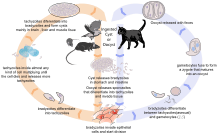
| Toxoplasmosis | Toxoplasma gondii (Apicomplexa) | Undercooked meat, cat feces, fetal infection in pregnancy |
| Babesiosis | Babesia spp. (Apicomplexa) | Deer tick (Ixodes scapularis) |
| Cryptosporidiosis | Cryptosporidium spp. (Apicomplexa) | Fecal contamination of food or water |
| Cyclosporiasis | Cyclospora cayetanensis (Apicomplexa) | Fecal contamination of food or water |
References
- Panno, Joseph (14 May 2014). The Cell: Evolution of the First Organism. Infobase Publishing. ISBN 9780816067367.
- Bertrand, Jean-Claude; Caumette, Pierre; Lebaron, Philippe; Matheron, Robert; Normand, Philippe; Sime-Ngando, Télesphore (2015-01-26). Environmental Microbiology: Fundamentals and Applications: Microbial Ecology. Springer. ISBN 9789401791182.
- Madigan, Michael T. (2012). Brock Biology of Microorganisms. Benjamin Cummings. ISBN 9780321649638.
- Yaeger, Robert G. (1996). Protozoa: Structure, Classification, Growth, and Development. NCBI. ISBN 9780963117212. Retrieved 2018-03-23.
- Goldfuß (1818). "Ueber die Classification der Zoophyten" [On the classification of zoophytes]. Isis, Oder, Encyclopädische Zeitung von Oken (in German). 2 (6): 1008–1019. From p. 1008: "Erste Klasse. Urthiere. Protozoa." (First class. Primordial animals. Protozoa.) [Note: each column of each page of this journal is numbered; there are two columns per page.]
- Cavalier-Smith, Thomas (1981). "Eukaryote kingdoms: seven or nine?". Bio Systems. 14 (3–4): 461–481. doi:10.1016/0303-2647(81)90050-2. PMID 7337818.
- Cavalier-Smith, Thomas (December 1993). "Kingdom protozoa and its 18 phyla". Microbiological Reviews. 57 (4): 953–994. PMC 372943. PMID 8302218.
- Cavalier-Smith, Thomas (23 June 2010). "Kingdoms Protozoa and Chromista and the eozoan root of the eukaryotic tree". Biology Letters. 6 (3): 342–345. doi:10.1098/rsbl.2009.0948. PMC 2880060. PMID 20031978.
- Ruggiero, Michael A.; Gordon, Dennis P.; Orrell, Thomas M.; Bailly, Nicolas; Bourgoin, Thierry; Brusca, Richard C.; Cavalier-Smith, Thomas; Guiry, Michael D.; Kirk, Paul M. (29 April 2015). "A Higher Level Classification of All Living Organisms". PLoS ONE. 10 (4): e0119248. Bibcode:2015PLoSO..1019248R. doi:10.1371/journal.pone.0119248. PMC 4418965. PMID 25923521.
- Goldfuß, Georg August (1820). Handbuch der Zoologie. Erste Abtheilung [Handbook of Zoology. First Part.] (in German). Nürnberg, (Germany): Johann Leonhard Schrag. pp. XI–XIV.
- Bailly, Anatole (1981-01-01). Abrégé du dictionnaire grec français. Paris: Hachette. ISBN 978-2010035289. OCLC 461974285.
- Bailly, Anatole. "Greek-french dictionary online". www.tabularium.be. Retrieved 2018-10-05.
- Hogg, John (1860). "On the distinctions of a plant and an animal, and on a fourth kingdom of nature". Edinburgh New Philosophical Journal. 2nd series. 12: 216–225.
- Scamardella, J. M. (December 1999). "Not plants or animals: a brief history of the origin of Kingdoms Protozoa, Protista and Protoctista". International Microbiology. 2 (4): 207–216. PMID 10943416.
- Copeland, Herbert F. (September–October 1947). "Progress Report on Basic Classification". The American Naturalist. 81 (800): 340–361. doi:10.1086/281531. JSTOR 2458229.
- Siebold (vol. 1); Stannius (vol. 2) (1848). Lehrbuch der vergleichenden Anatomie [Textbook of Comparative Anatomy] (in German). vol. 1: Wirbellose Thiere (Invertebrate animals). Berlin, (Germany): Veit & Co. p. 3. From p. 3: "Erste Hauptgruppe. Protozoa. Thiere, in welchen die verschiedenen Systeme der Organe nicht scharf ausgeschieden sind, und deren unregelmässige Form und einfache Organisation sich auf eine Zelle reduziren lassen." (First principal group. Protozoa. Animals, in which the different systems of organs are not sharply separated, and whose irregular form and simple organization can be reduced to one cell.)
- Dobell, C. (April 1951). "In memoriam Otto Bütschli (1848-1920) "architect of protozoology"". Isis; an International Review Devoted to the History of Science and Its Cultural Influences. 42 (127): 20–22. doi:10.1086/349230. PMID 14831973.
- Taylor, F. J. R. 'Max' (11 January 2003). "The collapse of the two-kingdom system, the rise of protistology and the founding of the International Society for Evolutionary Protistology (ISEP)". International Journal of Systematic and Evolutionary Microbiology. 53 (6): 1707–1714. doi:10.1099/ijs.0.02587-0. PMID 14657097.
- Whittaker, R. H. (10 January 1969). "New concepts of kingdoms or organisms. Evolutionary relations are better represented by new classifications than by the traditional two kingdoms". Science. 163 (3863): 150–160. Bibcode:1969Sci...163..150W. CiteSeerX 10.1.1.403.5430. doi:10.1126/science.163.3863.150. PMID 5762760.
- Margulis, Lynn (1974). "Five-Kingdom Classification and the Origin and Evolution of Cells". In Dobzhansky, Theodosius; Hecht, Max K.; Steere, William C. (eds.). Evolutionary Biology. Springer. pp. 45–78. doi:10.1007/978-1-4615-6944-2_2. ISBN 978-1-4615-6946-6.
- Cavalier-Smith, Thomas (August 1998). "A revised six-kingdom system of life". Biological Reviews. 73 (3): 203–266. doi:10.1111/j.1469-185X.1998.tb00030.x. PMID 9809012.
- "New President's Address". protozoa.uga.edu. Retrieved 1 May 2015.
- Singleton, Paul; Sainsbury, Diana (2001). Dictionary of microbiology and molecular biology. Wiley. ISBN 9780471941507.
- Gooday, A.J.; Aranda da Silva, A. P.; Pawlowski, J. (1 December 2011). "Xenophyophores (Rhizaria, Foraminifera) from the Nazaré Canyon (Portuguese margin, NE Atlantic)". Deep-Sea Research Part II: Topical Studies in Oceanography. 58 (24–25): 2401–2419. Bibcode:2011DSRII..58.2401G. doi:10.1016/j.dsr2.2011.04.005.
- Ghaffar, Abdul. "Blood and Tissue Protozoa". Microbiology and Immunology On-Line. Retrieved 2018-03-23.
- Mylnikov, Alexander P.; Weber, Felix; Jürgens, Klaus; Wylezich, Claudia (August 2015). "Massisteria marina has a sister: Massisteria voersi sp. nov., a rare species isolated from coastal waters of the Baltic Sea". European Journal of Protistology. 51 (4): 299–310. doi:10.1016/j.ejop.2015.05.002. PMID 26163290.
- Mitchell, Gary C.; Baker, J. H.; Sleigh, M. A. (1 May 1988). "Feeding of a freshwater flagellate, Bodo saltans, on diverse bacteria". The Journal of Protozoology. 35 (2): 219–222. doi:10.1111/j.1550-7408.1988.tb04327.x.
- Ghaffar, Abdul. "Blood and tissue Protozoa". Microbiology and Immunology On-Line. Retrieved 2018-03-23.
- "Trypanosoma brucei". parasite.org.au. Retrieved 2018-03-23.
- "Microscopy of Entamoeba histolytica". msu.edu. Retrieved 2016-08-21.
- Lehman, Don. "Diagnostic parasitology". University of Delaware. Retrieved 2018-03-23.
- Taylor, Bruce. "Paramecium caudatum". Encyclopedia of Life. Retrieved 2018-03-23.
- "Amoeba proteus | Microworld". www.arcella.nl. Retrieved 2016-08-21.
- "Noctiluca scintillans". University of Tasmania, Australia. 2011-11-30. Retrieved 2018-03-23.
- Sheehan, Kathy B. (2005). Seen and Unseen: Discovering the Microbes of Yellowstone. Falcon. ISBN 9780762730933.
- Post, F. J.; Borowitzka, L. J.; Borowitzka, M. A.; Mackay, B.; Moulton, T. (1983-09-01). "The protozoa of a Western Australian hypersaline lagoon". Hydrobiologia. 105 (1): 95–113. Bibcode:2004HyBio.524..167W. doi:10.1007/BF00025180. ISSN 0018-8158.
- "Termite gut microbes | NOLL LAB". www.kennethnoll.uconn.edu. Retrieved 2018-03-21.
- Williams, A. G.; Coleman, G. S. (1997). The Rumen Microbial Ecosystem. Springer, Dordrecht. pp. 73–139. doi:10.1007/978-94-009-1453-7_3. ISBN 9789401071499.
- Lee, John J.; Leedale, Gordon F.; Bradbury, Phyllis Clarke (25 May 2000). An illustrated guide to the protozoa: organisms traditionally referred to as protozoa, or newly discovered groups. Society of Protozoologists. p. 634.
- Wiser, Mark F. "Biochemistry of Plasmodium". The Wiser Page. Retrieved 2018-03-22.
- Nishitani, Goh; Nagai, Satoshi; Baba, Katsuhisa; Kiyokawa, Susumu; Kosaka, Yuki; Miyamura, Kazuyoshi; Nishikawa, Tetsuya; Sakurada, Kiyonari; Shinada, Akiyoshi (May 2010). "High-Level Congruence of Myrionecta rubra Prey and Dinophysis Species Plastid Identities as Revealed by Genetic Analyses of Isolates from Japanese Coastal Waters". Applied and Environmental Microbiology. 76 (9): 2791–2798. doi:10.1128/AEM.02566-09. PMC 2863437. PMID 20305031.
- Protozoa in biological research
- "Sex and Death in Protozoa". Cambridge University Press. Retrieved 2015-06-09.
- Bernstein H, Bernstein C (2013). Bernstein C, Bernstein H eds. Evolutionary Origin and Adaptive Function of Meiosis'. Meiosis. InTech. ISBN 978-953-51-1197-9
- Kudo, Richard R. (Richard Roksabro) (1954). Protozoology. MBLWHOI Library. Springfield, Ill., C. C. Thomas.
- Honigberg, B. M.; W. Balamuth; E. C. Bovee; J. O. Corliss; M. Gojdics; R. P. Hall; R. R. Kudo; N. D. Levine; A. R. Lobblich; J. Weiser (February 1964). "A Revised Classification of the Phylum Protozoa". Journal of Eukaryotic Microbiology. 11 (1): 7–20. doi:10.1111/j.1550-7408.1964.tb01715.x. PMID 14119564.
- Adl, Sina M.; Simpson, Alastair G. B.; Lane, Christopher E.; Lukeš, Julius; Bass, David; Bowser, Samuel S.; Brown, Matthew W.; Burki, Fabien; Dunthorn, Micah (2012-09-01). "The Revised Classification of Eukaryotes". Journal of Eukaryotic Microbiology. 59 (5): 429–514. doi:10.1111/j.1550-7408.2012.00644.x. PMC 3483872. PMID 23020233.
- Bartel, Rebecca; Oberhauser, Karen; De Roode, Jacob; Atizer, Sonya (February 2011). "Monarch butterfly migration and parasite transmission in eastern North America". Ecology. 92 (2): 342–351. doi:10.1890/10-0489.1. PMID 21618914.
- Leong, K. L. H.; M. A. Yoshimura; H. K. Kaya; H. Williams (January 1997). "Instar Susceptibility of the Monarch Butterfly (Danaus plexippus) to the Neogregarine Parasite, Ophryocystis elektroscirrha". Journal of Invertebrate Pathology. 69 (1): 79–83. CiteSeerX 10.1.1.494.9827. doi:10.1006/jipa.1996.4634. PMID 9028932.
- Usha mina, Pranav kumar (2014). Life science fundamental and practice part I.
Bibliography
- General
- Dogiel, V. A., revised by J.I. Poljanskij and E. M. Chejsin. General Protozoology, 2nd ed., Oxford University Press, 1965.
- Hausmann, K., N. Hulsmann. Protozoology. Thieme Verlag; New York, 1996.
- Kudo, R.R. Protozoology. Springfield, Illinois: C.C. Thomas, 1954; 4th ed.
- Manwell, R.D. Introduction to Protozoology, second revised edition, Dover Publications Inc., New York, 1968.
- Roger Anderson, O. Comparative protozoology: ecology, physiology, life history. Berlin [etc.]: Springer-Verlag, 1988.
- Sleigh, M. The Biology of Protozoa. E. Arnold: London, 1981.
- Identification
- Jahn,T.L.- Bovee, E.C. & Jahn, F.F. How to Know the Protozoa. Wm. C. Brown Publishers, Div. of McGraw Hill, Dubuque, Iowa, 1979; 2nd ed.
- Lee, J.J., Leedale, G.F. & Bradbury, P. An Illustrated Guide to the Protozoa. Lawrence, Kansas, U.S.A: Society of Protozoologists, 2000; 2nd ed.
- Patterson, D.J. Free-Living Freshwater Protozoa. A Colour Guide. Manson Publishing; London, 1996.
- Patterson, D.J., M.A. Burford. A Guide to the Protozoa of Marine Aquaculture Ponds. CSIRO Publishing, 2001.
- Morphology
- Harrison, F.W., Corliss, J.O. (ed.). 1991. Microscopic Anatomy of Invertebrates, vol. 1, Protozoa. New York: Wiley-Liss, 512 pp.
- Pitelka, D. R. 1963. Electron-Microscopic Structure of Protozoa. Pergamon Press, Oxford.
- Physiology and biochemistry
- Nisbet, B. 1984. Nutrition and feeding strategies in Protozoa. Croom Helm Publ., London, 280 pp.
- Coombs, G.H. & North, M. 1991. Biochemical protozoology. Taylor & Francis, London, Washington.
- Laybourn-Parry J. 1984. A Functional Biology of Free-Living Protozoa. Berkeley, California: University of California Press.
- Levandowski, M., S.H. Hutner (eds). 1979. Biochemistry and physiology of protozoa. Volumes 1, 2, and 3. Academic Press: New York, NY; 2nd ed.
- Sukhareva-Buell, N.N. 2003. Biologically active substances of protozoa. Dordrecht: Kluwer.
- Ecology
- Capriulo, G.M. (ed.). 1990. Ecology of Marine Protozoa. Oxford Univ. Press, New York.
- Darbyshire, J.F. (ed.). 1994. Soil Protozoa. CAB International: Wallingford, U.K. 2009 pp.
- Laybourn-Parry, J. 1992. Protozoan plankton ecology. Chapman & Hall, New York. 213 pp.
- Fenchel, T. 1987. Ecology of protozoan: The biology of free-living phagotrophic protists. Springer-Verlag, Berlin. 197 pp.
- Parasitology
- Kreier, J.P. (ed.). 1991-1995. Parasitic Protozoa, 2nd ed. 10 vols (1-3 coedited by Baker, J.R.). Academic Press, San Diego, California, .
- Methods
External links
| Wikispecies has information related to Protozoa |
- Chisholm, Hugh, ed. (1911). . Encyclopædia Britannica (11th ed.). Cambridge University Press.