Promyelocytic leukemia protein
Promyelocytic leukemia protein (PML) (also known as MYL, RNF71, PP8675 or TRIM19[5]) is the protein product of the PML gene. PML protein is a tumor suppressor protein required for the assembly of a number of nuclear structures, called PML-nuclear bodies, which form amongst the chromatin[5] of the cell nucleus. These nuclear bodies are present in mammalian nuclei, at about 1 to 30 per cell nucleus.[5] PML-NBs are known to have a number of regulatory cellular functions, including involvement in programmed cell death, genome stability, antiviral effects and controlling cell division.[5][6] PML mutation or loss, and the subsequent dysregulation of these processes, has been implicated in a variety of cancers.[5]
History
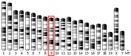
PML was poorly understood until described in the findings of Grignani et al in their 1996 study of patients with acute promylocytic leukemia (APL). It was found that the karyotype of 90% of APL patients included a reciprocal translocation, resulting in the fusion of the Retinoic Acid Receptor (RARalpha) gene of chromosome 17 and the PML gene of chromosome 15, which had not previously been characterized. The resultant PML/RARalpha oncofusion gene was shown to disturb normal PML and RARalpha function, thus inhibiting the terminal differentiation of blood precursor cells and allowing the maintenance of a reserve of undifferentiated cells for cancerous progression.[7] This implication of the PML gene in a pathological context led to a greater focus on the gene in future years.
Structure
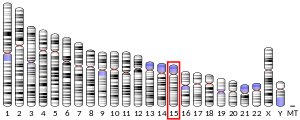
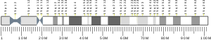
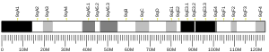
The PML gene is roughly 53 kilobase pairs in length and is located on the q arm of chromosome 15. It consists of 10 exons that are subject to shuffling through alternative splicing, yielding more than 15 known PML protein isoforms.[8][9] While the isoforms vary at their c-terminal domain, they all contain a TRIpartite motif encoded by the first three exons of the gene.[10] The TRIpartite motif consists of a zinc RING finger, two zinc binding domains, termed the B1 and B2 boxes, and an RBCC dimerization domain composed of two alpha helical coiled coil domains.[9]
The PML gene is under control at the transcriptional, translational and post translational control. The promoter region of the gene contains targets of Signal Transducer and Activator of Transcription (STATs), interferon regulatory factors, and p53 protein, indicating the intricacy of its involvement in cellular functions.[11] In addition to regulation through alternative splicing, the protein product is subject to post-translational modifications such as acetylation and phosphorylation. The c-terminus contains serine residues that are phosphorylated by casein kinases, and there are several tyrosine and threonine residues which can also be phosphorylation targets.[9] PML phosphorylation triggers further modification through the attachment of SUMO proteins to the RING domain by UBC9 SUMO-conjugating enzyme,[5] which occurs in a cell cycle dependent way. PML contains a SUMO-binding domain necessary for its interaction with other SUMOylated proteins such as itself and many others.[9] Both ubiquitination and SUMOylation of PML protein can trigger its degradation in the proteasome, thus providing a means of modulating PML protein lability within the cell.[11]
PML is translated in the cytoplasm of the cell, but its N-terminus contains a nuclear localization signal which causes its import to the nucleus.[9] Within the nucleus, sumoylated PML proteins multimerize with one another through interactions at the RBCC domain.This forms a ring-like structure that binds to the nuclear matrix, forming a PML-Nuclear body (PML-NB). The edge of the ring-like protein mutimer features protein threads that extend out from the ring and make contact with chromatin fibers.[5] This maintains the position of the PML-NBs within the nucleus, as well as the stability of the protein. When the chromatin is stressed, such as during apoptosis, the PML-NB becomes unstable and the PML bodies are redistributed into microstructures. These microstructures contain PML protein but not the many interacting proteins normally associated with PML-NBs.[5][12]
PML-NBs are not randomly distributed throughout the nucleus, but are found within the nucleus and are commonly associated with other nuclear bodies such as splicing speckles and nucleoi, as well as regions that are rich in genes and are actively being transcribed . Particularly, PML-NB have been shown to associate with genes such as the MHC I cluster of genes, as well as the p53 gene. The exact significance of this association is unclear, however evidence suggests that PML-NBs may influence transcription at these specific gene sites.[13]
Function
The PML-NBs have a wide array of functions, and a large role in cell regulation. They exert their wide range of actions through interactions with varying proteins localized to the PML-NBs. It is thought that the specific biochemical function performed by PML-NBs may be serving as an E3 ligase for the sumoylation of other proteins.[5] The true function, however, remains unclear, and several possible models have been proposed for PML-NB function, including nuclear storage of proteins, serving as a dock where other proteins accumulate to be post-translationally modified, direct involvement with transcription, and chromatin regulation.[5]
PML-NBs also play a role in transcriptional regulation. PML-NBs have been shown to increase the transcription of some genes, while repressing the transcription of other genes.[5] It has been suggested that the mechanism by which PML-NBs do this is via a chromatin-remodelling processes, although this is uncertain.[5]
Due to this apparent contradiction, it is possible that PML-NBs may be heterogeneous structures that have different functions based on their location within the nucleus, the proteins they interact with in a specific area of the nucleus, or the specific PML protein isoforms of which they are composed.
In addition to this regulation of transcription, observations of PML-NBs have strongly suggested that the protein complex plays a role in mediating DNA-damage responses. For example, the number and size of PML-NBs increases as the activities of DNA damage sensors ATM and ATR increase. The nuclear bodies localize to the site of DNA damage, where proteins associated with the repair of DNA and halting of the cell cycle then co-localize.[5][13] The functional purpose of the interaction between PML-NBs and DNA repair mechanisms remains unclear, but it seems unlikely that they have a role in repairing the DNA directly, due to the co-localization of DNA repair proteins and PML-NBs some time after the DNA has been damaged. Rather, it is thought that PML-NBs may regulate responses to DNA damage by acting as a storage site for proteins involved in DNA repair, regulating the repair directly, or mediating between DNA repair and checkpoint responses.[5] However, it is clear that PML-NBs play a role in mediating checkpoint responses, particularly in causing apoptosis.
PML plays an important role in both p53 dependent and p53 independent apoptotic pathways. PML activates p53 by recruiting the protein to a PML-NBs site and promoting its activation, while inhibiting regulators of the protein such as MDM2 or HAUSP.[5] In pathways that do not use p53 in inducing apoptosis, PML have been shown to interact with CHK2 and induce it to autophosphorylate to become active.[5] In addition to those two apoptotic pathways, Fas-induced apoptosis relies on the PML-NBs to release FLICE-Associated huge protein, which then localizes to the mitochondria to promote the activation of Caspase-8.[5]
Beyond apoptosis, other studies have implicated PML-NBs in cellular senescence, particularly its induction.[5] It has been shown to be involved with the formation of certain chromatin features of cells experiencing senescence, such as senescence-associated heterochromatin foci (SAHFs), which are believed to suppress the expression of growth-promoting factors and genes. The formation of these features is the result of histone chaperones, HIRA and ASF1, whose chromatin remodeling activities here are mediated by the PML-NBs. HIRA localizes to PML-NBs before any other interaction occurs with the DNA.[5]
Role in Cancer
Loss of function mutations of the PML protein, particularly resulting from the fusion of the PML gene with RARα gene in Acute promyelocytic leukemias, is implicated in several tumor suppressing apoptotic pathways, particularly those that rely on p53 as noted above.[5][14] Thus, the loss of PML function confers a cellular survival and proliferation advantage, impedes cellular senescence through loss of SAHFs, and puts a block on cellular differentiation.[14]
Both humans and mice have been found to demonstrate an increased propensity for tumor formation upon loss of PML function. PML disruption occurs in a wide variety of cancer types, and results in more metastatic tumors, and correspondingly poorer prognoses.[14] It is thought that, beyond the importance it plays in apoptotic roles, PML inactivation may cause cells to favor tumor progression by allowing the cell to accumulate additional genetic damage. Many proteins involved in genomic stability maintenance rely on the PML-NBs for targeting, and PML loss thus leads to a decrease in repair efficiency within the cell.[14]
Cell Cycle Role
PML-NB distribution and concentration changes as the cell moves through the cell cycle. In G0 phase, few sumoylated PML-NBs are present, but their numbers increase as the cell progresses through G1 to S to G2 stages. During the chromatin condensation occurring during mitosis, the desumoylation of PML causes the dissociation of many associated factors, and the PML proteins self aggregate to form a few, large, aggregates termed mitotic accumulations of PML proteins (MAPPs).[5] In addition to changes in numbers, PML-NBs also associate with different proteins over the lifetime of the cycle, and undergo significant biochemical changes in composition.[5]
During the S phase of the cell cycle, PML-NB complexes break apart as their chromatin scaffold changes during replication. The physical breaking of the PML-NBs into smaller fragments promotes the creation of more PML-NBs that exist in G2, however, the expression levels of the PML protein hasn't increased.[5] It is thought that this may serve to preserve the orientation of the chromatids with which the PML-NBs are associated, or monitor the integrity of replication forks.[5]
Antiviral functions
Transcription of PML is increased by the presence of interferon α/β and γ. It is thought that the increased numbers of PML-NBs that result from this increase in expression of the PML protein may result in the sequestering of viral proteins in the PML-NBs. Thus, the virus is unable to make use of them. The proteins held by PML-NBs are then sumoylated, inactivating the virions permanently.[6]
Interactions
Promyelocytic leukemia protein has been shown to interact with:
- ANKRD2,[15]
- CREB-binding protein,[16][17][18]
- Cyclin T1,[19]
- Death associated protein 6,[20][21][22][23]
- GATA2,[24]
- HDAC1,[25][26]
- HDAC3,[26]
- HHEX,[27]
- MAPK11,[28]
- MYB,[29]
- Mdm2,[30][31][32][33]
- Nerve Growth factor IB,[34]
- Nuclear receptor co-repressor 1,[25]
- Nuclear receptor co-repressor 2,[25][35]
- P53,[30][36][37]
- RPL11,[31]
- Retinoblastoma protein,[38]
- Retinoic acid receptor alpha,[16]
- SIN3A,[25]
- SKI protein,[25]
- STAT3,[39]
- Serum response factor[17] and
- Small ubiquitin-related modifier 1,[40][41]
- Sp1 transcription factor,[42]
- TOPBP1,[43]
- Thymine-DNA glycosylase,[44] and
- Zinc finger and BTB domain-containing protein 16.[45]
See also
- RING finger domain
References
- GRCh38: Ensembl release 89: ENSG00000140464 - Ensembl, May 2017
- GRCm38: Ensembl release 89: ENSMUSG00000036986 - Ensembl, May 2017
- "Human PubMed Reference:". National Center for Biotechnology Information, U.S. National Library of Medicine.
- "Mouse PubMed Reference:". National Center for Biotechnology Information, U.S. National Library of Medicine.
- Bernardi R, Pandolfi PP (December 2007). "Structure, dynamics and functions of promyelocytic leukaemia nuclear bodies". Nature Reviews. Molecular Cell Biology. 8 (12): 1006–16. doi:10.1038/nrm2277. PMID 17928811.
- Sahin U, Lallemand-Breitenbach V, de Thé H (November 2014). "PML nuclear bodies: regulation, function and therapeutic perspectives". The Journal of Pathology. 234 (3): 289–91. doi:10.1002/path.4426. PMID 25138686.
- Grignani F, Testa U, Rogaia D, Ferrucci PF, Samoggia P, Pinto A, Aldinucci D, Gelmetti V, Fagioli M, Alcalay M, Seeler J, Grignani F, Nicoletti I, Peschle C, Pelicci PG (September 1996). "Effects on differentiation by the promyelocytic leukemia PML/RARalpha protein depend on the fusion of the PML protein dimerization and RARalpha DNA binding domains". The EMBO Journal. 15 (18): 4949–58. doi:10.1002/j.1460-2075.1996.tb00875.x. PMC 452232. PMID 8890168.
- "PML promyelocytic leukemia [Homo sapiens (human)] - Gene - NCBI". www.ncbi.nlm.nih.gov. Retrieved 2016-12-06.
- Pearson M, Pelicci PG (2001). "PML interaction with p53 and its role in apoptosis and replicative senescence". Oncogene. 20 (49): 7250–6. doi:10.1038/sj.onc.1204856. PMID 11704853.
- Tang MK, Liang YJ, Chan JY, Wong SW, Chen E, Yao Y, Gan J, Xiao L, Leung HC, Kung HF, Wang H, Lee KK (2013-03-21). "Promyelocytic leukemia (PML) protein plays important roles in regulating cell adhesion, morphology, proliferation and migration". PLOS ONE. 8 (3): e59477. doi:10.1371/journal.pone.0059477. PMC 3605454. PMID 23555679.
- Zhou W, Bao S (March 2014). "PML-mediated signaling and its role in cancer stem cells". Oncogene. 33 (12): 1475–84. doi:10.1038/onc.2013.111. PMID 23563177.
- Ching RW, Dellaire G, Eskiw CH, Bazett-Jones DP (March 2005). "PML bodies: a meeting place for genomic loci?". Journal of Cell Science. 118 (Pt 5): 847–54. doi:10.1242/jcs.01700. PMID 15731002.
- Dellaire G, Bazett-Jones DP (September 2004). "PML nuclear bodies: dynamic sensors of DNA damage and cellular stress". BioEssays. 26 (9): 963–77. doi:10.1002/bies.20089. PMID 15351967.
- Gurrieri C, Capodieci P, Bernardi R, Scaglioni PP, Nafa K, Rush LJ, Verbel DA, Cordon-Cardo C, Pandolfi PP (February 2004). "Loss of the tumor suppressor PML in human cancers of multiple histologic origins". Journal of the National Cancer Institute. 96 (4): 269–79. doi:10.1093/jnci/djh043. PMID 14970276.
- Kojic S, Medeot E, Guccione E, Krmac H, Zara I, Martinelli V, Valle G, Faulkner G (May 2004). "The Ankrd2 protein, a link between the sarcomere and the nucleus in skeletal muscle". Journal of Molecular Biology. 339 (2): 313–25. doi:10.1016/j.jmb.2004.03.071. PMID 15136035.
- Zhong S, Delva L, Rachez C, Cenciarelli C, Gandini D, Zhang H, Kalantry S, Freedman LP, Pandolfi PP (November 1999). "A RA-dependent, tumour-growth suppressive transcription complex is the target of the PML-RARalpha and T18 oncoproteins". Nature Genetics. 23 (3): 287–95. doi:10.1038/15463. PMID 10610177.
- Matsuzaki K, Minami T, Tojo M, Honda Y, Saitoh N, Nagahiro S, Saya H, Nakao M (March 2003). "PML-nuclear bodies are involved in cellular serum response". Genes to Cells. 8 (3): 275–86. doi:10.1046/j.1365-2443.2003.00632.x. PMID 12622724.
- Doucas V, Tini M, Egan DA, Evans RM (March 1999). "Modulation of CREB binding protein function by the promyelocytic (PML) oncoprotein suggests a role for nuclear bodies in hormone signaling". Proceedings of the National Academy of Sciences of the United States of America. 96 (6): 2627–32. doi:10.1073/pnas.96.6.2627. PMC 15819. PMID 10077561.
- Marcello A, Ferrari A, Pellegrini V, Pegoraro G, Lusic M, Beltram F, Giacca M (May 2003). "Recruitment of human cyclin T1 to nuclear bodies through direct interaction with the PML protein". The EMBO Journal. 22 (9): 2156–66. doi:10.1093/emboj/cdg205. PMC 156077. PMID 12727882.
- Ishov AM, Sotnikov AG, Negorev D, Vladimirova OV, Neff N, Kamitani T, Yeh ET, Strauss JF, Maul GG (October 1999). "PML is critical for ND10 formation and recruits the PML-interacting protein daxx to this nuclear structure when modified by SUMO-1". The Journal of Cell Biology. 147 (2): 221–34. doi:10.1083/jcb.147.2.221. PMC 2174231. PMID 10525530.
- Li H, Leo C, Zhu J, Wu X, O'Neil J, Park EJ, Chen JD (March 2000). "Sequestration and inhibition of Daxx-mediated transcriptional repression by PML". Molecular and Cellular Biology. 20 (5): 1784–96. doi:10.1128/mcb.20.5.1784-1796.2000. PMC 85360. PMID 10669754.
- Lehembre F, Müller S, Pandolfi PP, Dejean A (January 2001). "Regulation of Pax3 transcriptional activity by SUMO-1-modified PML". Oncogene. 20 (1): 1–9. doi:10.1038/sj.onc.1204063. PMID 11244500.
- Zhong S, Salomoni P, Ronchetti S, Guo A, Ruggero D, Pandolfi PP (February 2000). "Promyelocytic leukemia protein (PML) and Daxx participate in a novel nuclear pathway for apoptosis". The Journal of Experimental Medicine. 191 (4): 631–40. doi:10.1084/jem.191.4.631. PMC 2195846. PMID 10684855.
- Tsuzuki S, Towatari M, Saito H, Enver T (September 2000). "Potentiation of GATA-2 activity through interactions with the promyelocytic leukemia protein (PML) and the t(15;17)-generated PML-retinoic acid receptor alpha oncoprotein". Molecular and Cellular Biology. 20 (17): 6276–86. doi:10.1128/mcb.20.17.6276-6286.2000. PMC 86102. PMID 10938104.
- Khan MM, Nomura T, Kim H, Kaul SC, Wadhwa R, Shinagawa T, Ichikawa-Iwata E, Zhong S, Pandolfi PP, Ishii S (June 2001). "Role of PML and PML-RARalpha in Mad-mediated transcriptional repression". Molecular Cell. 7 (6): 1233–43. doi:10.1016/s1097-2765(01)00257-x. PMID 11430826.
- Wu WS, Vallian S, Seto E, Yang WM, Edmondson D, Roth S, Chang KS (April 2001). "The growth suppressor PML represses transcription by functionally and physically interacting with histone deacetylases". Molecular and Cellular Biology. 21 (7): 2259–68. doi:10.1128/MCB.21.7.2259-2268.2001. PMC 86860. PMID 11259576.
- Topcu Z, Mack DL, Hromas RA, Borden KL (November 1999). "The promyelocytic leukemia protein PML interacts with the proline-rich homeodomain protein PRH: a RING may link hematopoiesis and growth control". Oncogene. 18 (50): 7091–100. doi:10.1038/sj.onc.1203201. PMID 10597310.
- Shin J, Park B, Cho S, Lee S, Kim Y, Lee SO, Cho K, Lee S, Jin BS, Ahn JH, Choi EJ, Ahn K (September 2004). "Promyelocytic leukemia is a direct inhibitor of SAPK2/p38 mitogen-activated protein kinase". The Journal of Biological Chemistry. 279 (39): 40994–1003. doi:10.1074/jbc.M407369200. PMID 15273249.
- Dahle Ø, Bakke O, Gabrielsen OS (July 2004). "c-Myb associates with PML in nuclear bodies in hematopoietic cells". Experimental Cell Research. 297 (1): 118–26. doi:10.1016/j.yexcr.2004.03.014. PMID 15194430.
- Kurki S, Latonen L, Laiho M (October 2003). "Cellular stress and DNA damage invoke temporally distinct Mdm2, p53 and PML complexes and damage-specific nuclear relocalization". Journal of Cell Science. 116 (Pt 19): 3917–25. doi:10.1242/jcs.00714. PMID 12915590.
- Bernardi R, Scaglioni PP, Bergmann S, Horn HF, Vousden KH, Pandolfi PP (July 2004). "PML regulates p53 stability by sequestering Mdm2 to the nucleolus". Nature Cell Biology. 6 (7): 665–72. doi:10.1038/ncb1147. PMID 15195100.
- Zhu H, Wu L, Maki CG (December 2003). "MDM2 and promyelocytic leukemia antagonize each other through their direct interaction with p53". The Journal of Biological Chemistry. 278 (49): 49286–92. doi:10.1074/jbc.M308302200. PMID 14507915.
- Wei X, Yu ZK, Ramalingam A, Grossman SR, Yu JH, Bloch DB, Maki CG (August 2003). "Physical and functional interactions between PML and MDM2". The Journal of Biological Chemistry. 278 (31): 29288–97. doi:10.1074/jbc.M212215200. PMID 12759344.
- Wu WS, Xu ZX, Ran R, Meng F, Chang KS (May 2002). "Promyelocytic leukemia protein PML inhibits Nur77-mediated transcription through specific functional interactions". Oncogene. 21 (24): 3925–33. doi:10.1038/sj.onc.1205491. PMID 12032831.
- Hong SH, Yang Z, Privalsky ML (November 2001). "Arsenic trioxide is a potent inhibitor of the interaction of SMRT corepressor with Its transcription factor partners, including the PML-retinoic acid receptor alpha oncoprotein found in human acute promyelocytic leukemia". Molecular and Cellular Biology. 21 (21): 7172–82. doi:10.1128/MCB.21.21.7172-7182.2001. PMC 99892. PMID 11585900.
- Fogal V, Gostissa M, Sandy P, Zacchi P, Sternsdorf T, Jensen K, Pandolfi PP, Will H, Schneider C, Del Sal G (November 2000). "Regulation of p53 activity in nuclear bodies by a specific PML isoform". The EMBO Journal. 19 (22): 6185–95. doi:10.1093/emboj/19.22.6185. PMC 305840. PMID 11080164.
- Guo A, Salomoni P, Luo J, Shih A, Zhong S, Gu W, Pandolfi PP (October 2000). "The function of PML in p53-dependent apoptosis". Nature Cell Biology. 2 (10): 730–6. doi:10.1038/35036365. PMID 11025664.
- Alcalay M, Tomassoni L, Colombo E, Stoldt S, Grignani F, Fagioli M, Szekely L, Helin K, Pelicci PG (February 1998). "The promyelocytic leukemia gene product (PML) forms stable complexes with the retinoblastoma protein". Molecular and Cellular Biology. 18 (2): 1084–93. doi:10.1128/mcb.18.2.1084. PMC 108821. PMID 9448006.
- Kawasaki A, Matsumura I, Kataoka Y, Takigawa E, Nakajima K, Kanakura Y (May 2003). "Opposing effects of PML and PML/RAR alpha on STAT3 activity". Blood. 101 (9): 3668–73. doi:10.1182/blood-2002-08-2474. PMID 12506013.
- Lin DY, Shih HM (July 2002). "Essential role of the 58-kDa microspherule protein in the modulation of Daxx-dependent transcriptional repression as revealed by nucleolar sequestration". The Journal of Biological Chemistry. 277 (28): 25446–56. doi:10.1074/jbc.M200633200. PMID 11948183.
- Kamitani T, Nguyen HP, Kito K, Fukuda-Kamitani T, Yeh ET (February 1998). "Covalent modification of PML by the sentrin family of ubiquitin-like proteins". The Journal of Biological Chemistry. 273 (6): 3117–20. doi:10.1074/jbc.273.6.3117. PMID 9452416.
- Vallian S, Chin KV, Chang KS (December 1998). "The promyelocytic leukemia protein interacts with Sp1 and inhibits its transactivation of the epidermal growth factor receptor promoter". Molecular and Cellular Biology. 18 (12): 7147–56. doi:10.1128/MCB.18.12.7147. PMC 109296. PMID 9819401.
- Xu ZX, Timanova-Atanasova A, Zhao RX, Chang KS (June 2003). "PML colocalizes with and stabilizes the DNA damage response protein TopBP1". Molecular and Cellular Biology. 23 (12): 4247–56. doi:10.1128/mcb.23.12.4247-4256.2003. PMC 156140. PMID 12773567.
- Takahashi H, Hatakeyama S, Saitoh H, Nakayama KI (February 2005). "Noncovalent SUMO-1 binding activity of thymine DNA glycosylase (TDG) is required for its SUMO-1 modification and colocalization with the promyelocytic leukemia protein". The Journal of Biological Chemistry. 280 (7): 5611–21. doi:10.1074/jbc.M408130200. PMID 15569683.
- Koken MH, Reid A, Quignon F, Chelbi-Alix MK, Davies JM, Kabarowski JH, Zhu J, Dong S, Chen S, Chen Z, Tan CC, Licht J, Waxman S, de Thé H, Zelent A (September 1997). "Leukemia-associated retinoic acid receptor alpha fusion partners, PML and PLZF, heterodimerize and colocalize to nuclear bodies". Proceedings of the National Academy of Sciences of the United States of America. 94 (19): 10255–60. doi:10.1073/pnas.94.19.10255. PMC 23349. PMID 9294197.
- Miki T, Zhao Z, Lee CC (July 2016). "Interactive Organization of the Circadian Core Regulators PER2, BMAL1, CLOCK and PML". Scientific Reports. 6: 29174. doi:10.1038/srep29174. PMC 4935866. PMID 27383066.
Further reading
- Zhong S, Salomoni P, Pandolfi PP (May 2000). "The transcriptional role of PML and the nuclear body". Nature Cell Biology. 2 (5): E85–90. doi:10.1038/35010583. PMID 10806494.
- Jensen K, Shiels C, Freemont PS (October 2001). "PML protein isoforms and the RBCC/TRIM motif". Oncogene. 20 (49): 7223–33. doi:10.1038/sj.onc.1204765. PMID 11704850.
- Pearson M, Pelicci PG (October 2001). "PML interaction with p53 and its role in apoptosis and replicative senescence". Oncogene. 20 (49): 7250–6. doi:10.1038/sj.onc.1204856. PMID 11704853.
- Salomoni P, Pandolfi PP (January 2002). "The role of PML in tumor suppression". Cell. 108 (2): 165–70. doi:10.1016/S0092-8674(02)00626-8. PMID 11832207.
- Combes R, Balls M, Bansil L, Barratt M, Bell D, Botham P, Broadhead C, Clothier R, George E, Fentem J, Jackson M, Indans I, Loizu G, Navaratnam V, Pentreath V, Phillips B, Stemplewski H, Stewart J (2002). "An assessment of progress in the use of alternatives in toxicity testing since the publication of the report of the second FRAME Toxicity Committee (1991)". Alternatives to Laboratory Animals. 30 (4): 365–406. doi:10.1177/026119290203000403. PMID 12234245.
- Bernardi R, Pandolfi PP (December 2003). "Role of PML and the PML-nuclear body in the control of programmed cell death". Oncogene. 22 (56): 9048–57. doi:10.1038/sj.onc.1207106. PMID 14663483.
- Beez S, Demmer P, Puccetti E (2012). "Targeting the acute promyelocytic leukemia-associated fusion proteins PML/RARα and PLZF/RARα with interfering peptides". PLOS ONE. 7 (11): e48636. doi:10.1371/journal.pone.0048636. PMC 3494703. PMID 23152790.
External links
- PML+protein,+human at the US National Library of Medicine Medical Subject Headings (MeSH)
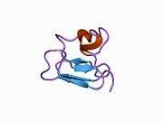
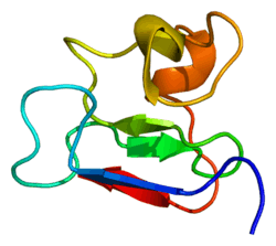
PDB gallery | |
|---|---|
|