Accessory auricle
An accessory auricle is considered a developmental anomaly resulting from the persistence of a structure which variably recapitulates the normal external ear.
| Accessory auricle | |
|---|---|
 | |
| Specialty | Medical genetics |
Signs and symptoms
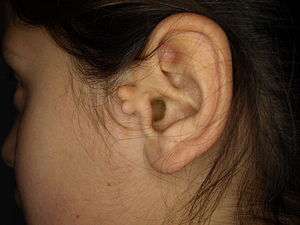
The general presentation is of a skin-covered nodule, papule, or nodule of the skin surface, usually immediately anterior to the auricle. However, it may be anywhere within the periauricular tissues. Bilateral presentation can be seen.[1][2][3][4][5]
Genetics
A study of a family with 11 affected individuals showed the phenotype was inherited in an autosomal dominant manner.[6]
Diagnosis
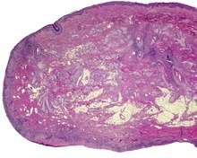
The lesions presents as a nodule or papule, either sessile or pedunculated. They may be soft or have a cartilaginous structure. By histologic examination, it is a recapitulation of normal external auricle. There will be skin, cartilaginous structures, and cartilage (although the later is not seen in all variants of this disorder).[3][4] Some investigators believe that the tragus is the only hillock which is derived from the first branchial arch. This is clearly suggestive that true cases of Accessory Auricle represent a true duplication of the hillocks that were part of the second branchial arch.[7] The second ear appears as a mirror image folded forward and lying on the posterior cheek.
Differential diagnosis
These structures are distinctly different from squamous papilloma and benign teratoma.
Classification
The several components or degrees of development range from an ear tag, preauricular appendage, preauricular tag, or accessory tragus, to supernumerary ears or polyotia.[8] It is a relatively common congenital anomaly of the first branchial arch or second branchial arches. Other anomalies may be present concurrently, including cleft palate, cleft lip, or mandibular hypoplasia. There is a known association with Goldenhar syndrome (oculo-auriculo-vertebral syndrome)[9] and with Wildervanck syndrome.[10][11][12] There may also be an association with congenital cartilaginous rest of the neck.
Management
Simple surgical excision is curative.[13] The recommended treatment is that the skin is peeled off the extra-auricular tissue and protruding cartilage remnants are trimmed.[14] Normal appearance is achieved in majority of cases. The reconstruction successful in true cases of accessory auricle, as it also is in individuals with auricular appendages.[15][16]
Epidemiology
These lesions usually present in neonates, although they may not come to clinical attention until adulthood (for cosmetic reasons). There is no gender predilection. They are present in approximately 3-6 per 1000 live births.[17]
References
- Cosman, B. C. (1993). "Bilateral accessory tragus". Cutis. 51 (3): 199–200. PMID 8444054.
- "Preauricular sinus and accessory auricle. (Photoclinic)." Consultant Feb. 2002: 256+. Health Reference Center Academic. Web. 2 Nov. 2011.
- Jansen, T.; Romiti, R.; Altmeyer, P. (2000). "Accessory tragus: Report of two cases and review of the literature". Pediatric Dermatology. 17 (5): 391–394. doi:10.1046/j.1525-1470.2000.017005391.x. PMID 11085670.
- Brownstein, M. H.; Wanger, N.; Helwig, E. B. (1971). "Accessory tragi". Archives of Dermatology. 104 (6): 625–631. doi:10.1001/archderm.1971.04000240049006. PMID 5131708.
- Hodges FR; Sahouria JJ; Wood AJ (2006). "Accessory tragus: A report of 2 cases". Journal of Dentistry for Children. 73 (1): 42–4. PMID 16734313.
- Yang, Y; et al. (2006). "A locus for autosomal dominant accessory auricular anomaly maps to 14q11.2–q12". Human Genetics. 120 (1): 144–147. doi:10.1007/s00439-006-0206-1. PMID 16775710.
- Stevenson, Roger E.; Hall, Judith G. (2005). Human Malformations and Related Anomalies (2nd ed.). Oxford University Press. pp. 339–340. ISBN 978-0199748082.
- Lam, J.; Dohil, M. (2007). "Multiple Accessory Tragi and Hemifacial Microsomia". Pediatric Dermatology. 24 (6): 657–658. doi:10.1111/j.1525-1470.2007.00560.x. PMID 18035991.
- Konaş, E.; Canter, H. I.; Mavili, M. E. (2006). "Goldenhar complex with atypical associated anomalies: Is the spectrum still widening?". Journal of Craniofacial Surgery. 17 (4): 669–672. doi:10.1097/00001665-200607000-00011. PMID 16877912.
- Tadini, G.; Cambiaghi, S.; Scarabelli, G.; Brusasco, A.; Vigo, P. (1993). "Familial occurrence of isolated accessory tragi". Pediatric Dermatology. 10 (1): 26–28. doi:10.1111/j.1525-1470.1993.tb00006.x. PMID 8493161.
- Gao, J. Z.; Chen, Y. M.; Gao, Y. P. (1990). "A survey of accessory auricle anomaly. Pedigree analysis of seven cases". Archives of Otolaryngology–Head & Neck Surgery. 116 (10): 1194–1196. doi:10.1001/archotol.1990.01870100088019. PMID 2206506.
- Resnick, K. I.; Soltani, K.; Bernstein, J. E.; Fathizadeh, A. (1981). "Accessory tragi and associated syndromes involving the first branchial arch". Journal of Dermatologic Surgery and Oncology. 7 (1): 39–41. doi:10.1111/j.1524-4725.1981.tb00591.x. PMID 7204730.
- Pan, B.; Qie, S.; Zhao, Y.; Tang, X.; Lin, L.; Yang, Q.; Zhuang, H.; Jiang, H. (2010). "Surgical management of polyotia". Journal of Plastic, Reconstructive & Aesthetic Surgery. 63 (8): 1283–1288. doi:10.1016/j.bjps.2009.06.037. PMID 19617017.
- Scott-Brown's Otorhinolaryngology (7th ed.). Hodder Arnold. 2016-06-15. p. 969. ISBN 978 0 340 808 931.
- Ku, PK; Tong, MC; Yue, V (1998). "Polyotia- a rare external ear anomaly". International Journal of Pediatric Otorhinolaryngology. 46 (1–2): 117. doi:10.1016/S0165-5876(98)00152-9.
- Bendor-Samuel, RL; Tung, TC; Chen, YR (1995). "Polyotia". Annals of Plastic Surgery. 34 (6): 650–2. doi:10.1097/00000637-199506000-00015. PMID 7661545.
- Rapini, Ronald P.; Bolognia, Jean L.; Jorizzo, Joseph L. (2007). Dermatology: 2-Volume Set. St. Louis: Mosby. p. 894. ISBN 978-1-4160-2999-1.
Further reading
Lester D. R. Thompson; Bruce M Wenig (2011). Diagnostic Pathology: Head and Neck. Hagerstown, MD: Lippincott Williams & Wilkins. pp. 7:2–3. ISBN 978-1-931884-61-7.