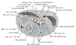
Posterior compartment of the forearm
The posterior compartment of the forearm (or extensor compartment)[2] contains twelve muscles which are chiefly responsible for extension of the wrist and digits, and supination of the forearm. It is separated from the anterior compartment by the interosseous membrane between the radius and ulna.
| Posterior compartment of the forearm | |
|---|---|
 Extensor compartment of the forearm and hand | |
| Details | |
| System | Musculoskeletal system |
| Artery | radial artery, radial recurrent artery, profunda brachii, posterior interosseous artery |
| Nerve | radial nerve,[1] posterior interosseous nerve |
| Identifiers | |
| Latin | compartimentum antebrachii posterius |
| TA | A04.6.01.007 |
| FMA | 38411 |
| Anatomical terminology | |
Structure
Muscles
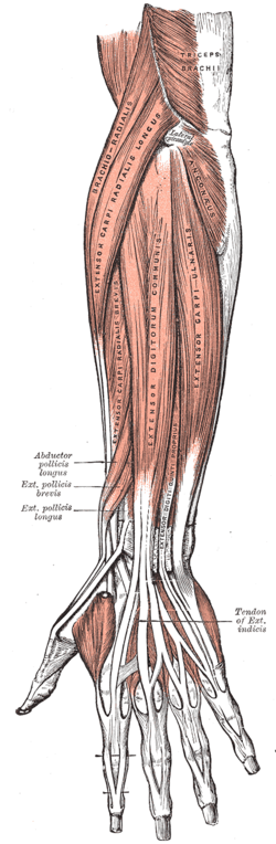
There are generally twelve muscles in the posterior compartment of the forearm, which can be further divided into a superficial, intermediate, and deep layer. Most of the muscles in the superficial and the intermediate layers share a common origin which is the outer part of the elbow, the lateral epicondyle of humerus. The deep muscles arise from the distal part of the ulna and the surrounding interosseous membrane.
The brachioradialis, flexor of the elbow, is unusual in that it is located in the posterior compartment, but it is actually a muscle of flexor / anterior compartment of the forearm. The anconeus, assisting in extension of the elbow joint, is by some considered part of the posterior compartment of the arm.
The majority of muscles found in the posterior compartment are extrinsic, meaning its origin has some distance from the part that it moves. The brachioradialis and the anconeus are considered intrinsic muscles because they both arise within the forearm and they both move the forearm.
| Level | Muscle | Extrinsic/Intrinsic | Innervation[3] |
| superficial | brachioradialis | intrinsic | radial nerve |
| superficial | extensor carpi radialis longus | extrinsic | radial nerve |
| superficial | extensor carpi radialis brevis | extrinsic | radial nerve (deep branch) |
| superficial | extensor carpi ulnaris | extrinsic | radial nerve (as posterior interosseous nerve) |
| superficial | anconeus | intrinsic | radial nerve |
| intermediate | extensor digitorum | extrinsic | radial nerve (as posterior interosseous nerve) |
| intermediate | extensor digiti minimi | extrinsic | radial nerve (as posterior interosseous nerve) |
| deep | abductor pollicis longus | extrinsic | radial nerve (as posterior interosseous nerve) |
| deep | extensor pollicis longus | extrinsic | radial nerve (as posterior interosseous nerve) |
| deep | extensor pollicis brevis | extrinsic | radial nerve (as posterior interosseous nerve) |
| deep | extensor indicis | extrinsic | radial nerve (as posterior interosseous nerve) |
| deep | supinator | intrinsic | radial nerve (deep branch) |
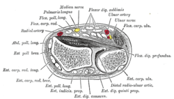
Extensor tendon compartments
Extensor tendons pass through the extensor retinaculum at wrist joint in 6 synovial sheaths, also referred to compartments.[3]
The supinator and the anconeus are the two muscles in the posterior compartment of the forearm that do not pass through wrist extensor compartments.[3]
.PNG)
- The first compartment locating the most radial is occupied by the extensor pollicis brevis and the abductor pollicis longus to insert to the thumb.
- The second compartment is occupied by the two radial wrist extensors, the extensor carpi radialis longus and the extensor carpi radialis brevis.
- The third compartment exclusively accommodates the extensor pollicis longus, which hooks around Lister's tubercle of radius and inserts to the thumb.
- The fourth compartment is the largest of all. It is occupied by the extensors of the digits, the extensor digitorum communis and the extensor indicis proprius. The extensor indicis proprius usually runs and inserts onto the ulnar side of the extensor digitorum communis of the index finger.[4]
- The fifth compartment is occupied by the extensor digiti minimi, the extensor of the little finger.
- The extensor carpi ulnaris passes through the sixth compartment to insert to the base of the fifth metacarpal bone.
Innervation
The muscles of the posterior compartment of the forearm are innervated by the radial nerve and its branches.[3] The radial nerve arises from the posterior cord of the plexus. The somatomotor fibers of the radial nerve branch from the main radial nerve at the level of the radial groove of the humerus.
Development
In the early stage of development, the extensor precursor divides into 3 layers namely, superficial layer, radial layer and deep layer.[5][6] The superficial group develops to become the extensor digitorum communis, the extensor carpi ulnaris and the extensor digiti minimi. The radial layer forms the extensor carpi radialis longus, the extensor carpi radialis brevis and the brachioradialis. The deep layer differentiates to become the abductor pollicis longus, the extensor pollicis longus and the extensor pollicis brevis.
| Phylogenetic origin[6] | Embryologic origin[6] | Representatives in humans[6] |
|---|---|---|
| Brachioantebrachial group | Radial layer | Brachioradialis |
| Extensor carpi radialis longus | ||
| Extensor carpi radialis brevis | ||
| Supinator | ||
| Superficial layer | Extensor digitorum communis | |
| Extensor carpi ulnaris | ||
| Extensor digiti minimi | ||
| Antebrachiomanual group | Deep layer (radial) | Extensor pollicis brevis |
| Abductor pollicis longus | ||
| Deep layer (ulnar) | Extensor pollicis longus | |
| Extensor indicis proprius | ||
| Extensor digitorum brevis manus[7] | ||
| Manual group | - | - |
Variations
The deep layer of the precursor extensor mass is known to be phylogenetically unstable and is undergoing evolution as high variability is seen in non-human primates.[5][8][9][10] In humans, anomalous or additional muscles can be seen in small portion of population.
Anomalous muscles in human extensor compartment are listed as follow:
- Extensor medii proprius
- Extensor indicis et medii communis
- Extensor pollicis et indicis communis
- Extensor carpi radialis tertius
- Extensor digitorum brevis manus
Clinical significance
Tennis elbow
Tennis elbow or lateral epicondylitis is a chronic or an acute inflammation of the tendons that arise from the outer part of the elbow. The affected tendons are the tendons of extensor muscles which originate from the lateral epicondyle of humerus. It is caused by the repetitive movements and overuse. It damages the tendons which results in pain and tenderness on the outer part of the elbow.[11]
De Quervain's syndrome
De Quervain's syndrome is a medical condition when the synovial sheath surrounding tendons in the first extensor tendon compartment becomes inflamed, so called tenosynovitis.[12] The tendons of the abductor pollicis longus and the extensor pollicis brevis run narrower due to the thickening of the synovial sheath, which causes pain when extending and moving the thumb outward.[13]
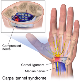
Carpal tunnel syndrome
Carpal tunnel syndrome is a condition when the median nerve is compressed as it passes through the carpal tunnel, which causes pain, numbness and tingling.
Fourth compartment syndrome
The presence of an additional tendon may result in a condition called fourth compartment syndrome.[14] Supernumerary tendons are common in the fourth extensor tendon compartment. Supernumerary tendons can refer to the additional tendons of normal structures or tendons of rare anatomical variants such as the extensor medii proprius or the extensor digitorum brevis manus. The increased pressure in the synovial sheath is known to directly or indirectly compress the posterior interosseous nerve of radial nerve.[14] Also, the extra pressure causes synovitis which results in pain in the dorsal part of the wrist.[15]
In other animals
In the superfamily hominoidea or apes, configurations of the muscles of the posterior compartment of the forearm share similar characteristics. However, the anconeus is usually not present in the hylobates (gibbons).[19] Also, the extensor pollicis brevis is only present in the genus homo (humans) and the genus hylobates because the extensor pollicis brevis and the abductor pollicis longus exist as a single muscle in other genera.[20]
Additional images
References
- lesson5musofpostforearm at The Anatomy Lesson by Wesley Norman (Georgetown University)
- "Dissector Answers - Forearm & Wrist". Retrieved 2008-01-17.
- Netter, Frank. H. (2014-11-14). Atlas of Human Anatomy, 6th Edition | Frank Netter |. store.elsevier.com. ISBN 9781455704187. Retrieved 2015-06-16.
- "Anatomy of the human body". archive.org. Retrieved 2015-06-16.
- Straus WL (1941) The phylogeny of the human forearm extensors. Hum Biol 13, 23–50.
- Cavdar, S.; Dogan, T.; Bayramiçli, M.; Sehirli, U.; Yüksel, M. (Jan 1998). "An unusual variation of extensor digitorum brevis manus: a case report and literature review". The Journal of Hand Surgery. 23 (1): 173–177. doi:10.1016/S0363-5023(98)80108-1. ISSN 0363-5023. PMID 9523974.
- Souter, W. A. (September 1, 1966). "The extensor digitorum brevis manus". British Journal of Surgery. 53 (9): 821–823. doi:10.1002/bjs.1800530923. ISSN 1365-2168. PMID 5911774.
- Wood, John (1866). "Variations in Human Myology Observed during the Winter Session of 1865-66 at King's College, London". Proceedings of the Royal Society of London. 15: 229–244. Bibcode:1866RSPS...15..229W. doi:10.1098/rspl.1866.0054. ISSN 0370-1662.
- Yoshida, Y.; Yasutaka, S.; Seki, Y. (August 1984). "[A study on the extensor digitorum brevis manus muscle in man]". Kaibogaku Zasshi. 59 (4): 313–321. ISSN 0022-7722. PMID 6524291.
- Čihák, Radomír (1972). Ontogenesis of the Skeleton and Intrinsic Muscles of the Human Hand and Foot. Springer Science & Business Media. ISBN 9783662090817.
- "Tennis Elbow (Lateral Epicondylitis) -OrthoInfo - AAOS". orthoinfo.aaos.org. Retrieved 2015-06-16.
- Ilyas, Asif M.; Ilyas, Asif; Ast, Michael; Schaffer, Alyssa A.; Thoder, Joseph (Dec 2007). "De quervain tenosynovitis of the wrist". The Journal of the American Academy of Orthopaedic Surgeons. 15 (12): 757–764. doi:10.5435/00124635-200712000-00009. ISSN 1067-151X. PMID 18063716.
- "de Quervain Syndrome". Handcare.org. Retrieved 2015-06-16.
- Hoffman, J.; Ellison, M. R. (Mar 1987). "Extensor digitorum brevis manus in the nondominant hand of two brothers". The Journal of Hand Surgery. 12 (2): 293–294. doi:10.1016/s0363-5023(87)80293-9. ISSN 0363-5023. PMID 3559091.
- Tan, S. T.; Smith, P. J. (May 1999). "Anomalous extensor muscles of the hand: a review". The Journal of Hand Surgery. 24 (3): 449–455. doi:10.1053/jhsu.1999.0449. ISSN 0363-5023. PMID 10357521.
- Ogura, T.; Inoue, H.; Tanabe, G. (Jan 1987). "Anatomic and clinical studies of the extensor digitorum brevis manus". The Journal of Hand Surgery. 12 (1): 100–107. doi:10.1016/s0363-5023(87)80171-5. ISSN 0363-5023. PMID 3805622.
- Patel, M. R.; Desai, S. S.; Bassini-Lipson, L.; Namba, T.; Sahoo, J. (Jul 1989). "Painful extensor digitorum brevis manus muscle". The Journal of Hand Surgery. 14 (4): 674–678. doi:10.1016/0363-5023(89)90190-1. ISSN 0363-5023. PMID 2754202.
- Anderson, M. W.; Benedetti, P.; Walter, J.; Steinberg, D. R. (Jun 1995). "MR appearance of the extensor digitorum manus brevis muscle: a pseudotumor of the hand". AJR. American Journal of Roentgenology. 164 (6): 1477–1479. doi:10.2214/ajr.164.6.7754896. ISSN 0361-803X. PMID 7754896.
- Diogo, R.; Abdala, V.; Lonergan, N.; Wood, B. A. (Oct 2008). "From fish to modern humans--comparative anatomy, homologies and evolution of the head and neck musculature". Journal of Anatomy. 213 (4): 391–424. doi:10.1111/j.1469-7580.2008.00953.x. ISSN 1469-7580. PMC 2644766. PMID 18657257.
- Aversi-Ferreira, T. A.; Diogo, R.; Potau, J. M.; Bello, G.; Pastor, J. F.; Aziz, M. Ashraf (Dec 2010). "Comparative anatomical study of the forearm extensor muscles of Cebus libidinosus (Rylands et al., 2000; Primates, Cebidae), modern humans, and other primates, with comments on primate evolution, phylogeny, and manipulatory behavior". Anatomical Record. 293 (12): 2056–2070. doi:10.1002/ar.21275. ISSN 1932-8494. PMID 21082733.
| Authority control |
|---|