Posterior clinoid processes
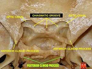
In the sphenoid bone, the anterior boundary of the sella turcica is completed by two small eminences, one on either side, called the anterior clinoid processes, while the posterior boundary is formed by a square-shaped plate of bone, the dorsum sellæ, ending at its superior angles in two tubercles, the posterior clinoid processes, the size and form of which vary considerably in different individuals. The posterior clinoid processes deepen the sella turcica, and give attachment to the tentorium cerebelli.
| Posterior clinoid processes | |
|---|---|
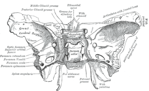 Sphenoid bone. Superior view. (Posterior clinoid process labeled at upper left.) | |
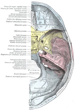 Base of the skull. Upper surface. (Caption for posterior clinoid process visible at center left. Sphenoid bone is yellow.) | |
| Details | |
| Identifiers | |
| Latin | Processus clinoideus posterior |
| TA | A02.1.05.011 |
| FMA | 54696 |
| Anatomical terms of bone | |
The petroclinoid ligament
The petroclinoid ligament is a fold of dura matter. It extends between the posterior clinoid process and anterior clinoid process and the petrosal part of the temporal bone of the skull. There are two separate bands of the ligament; named the anterior and posterior petroclinoid ligaments respectively. The anterior petroclinoid ligament is considered to be an extension of the tentorium cerebelli and the posterior petroclinoid ligament arises from the posteromedial extensions of the tentorial notch. The anterior and posterior petroclinoid ligaments are bands composed of collagen and elastic fibres that are densely packed in fascicles [1]
Their function:
The anterior petroclinoid ligament acts to laterally limit the superior wall of the cavernous sinus. The posterior petroclinoid ligament limits the posterior wall of the cavernous sinus. The angle between the two ligaments varies from 20 to 55 degrees.[2][3]
Anatomical Relations and Clinical significance:
The posterior petroclinoid ligament is in close proximity to the oculomotor nerve. During head trauma, it acts as a fulcrum following the downward displacement of the brainstem. This can cause injury to the pupillomotor fibres of the oculomotor nerve, consequently leading to internal opthalmoplegia [4]
The petroclinoid ligament attaches across the notch at the petrosphenoid junction. This forms a foramen, and within this lies the abducens nerve. The abducens nerve travels inferiorly to the petroclinoid ligament [5]
Ossification
The petroclinoid ligament could calcify. An ossified form of the ligament may create a syndrome, and this can be seen on a radiograph. The ossified ligament is a typical anatomical anomaly.[6][7]
Etymology
Clinoid likely comes from the Greek root klinein or the Latin clinare, both meaning "sloped" as in "inclined."
References
- J. Skrzat, J. Walocha, J.K. Jaworek, I. Mróz (23 November 2006). "The clinical significance of the petroclinoid ligament" (PDF). Via Medica. 66: 39–43.CS1 maint: multiple names: authors list (link)
- Lang, Johannes (1995). Skull base and related structure. Atlas of clinical anatomy. New York: Shattauer, Stutgart.
- Moore KL, Dalley AF (1999). Clinically oriented anatomy. 4th Edition. Philadelphia: Lippincott Williams & Wilkins.
- Nagaseki Y, Shimizu T, Kakizawa T, Fukamachi A, Nukui H (1989). "Primary internal ophthalmoplegia due to head injury". Acta Neurochir (Wien). 97 (3–4): 117–122. doi:10.1007/BF01772821. PMID 2718803.
- Piffer CR, Zorzetto NL (1980). "Course and relations of the abducens nerve". Anat Anz. 147: 42–46.
- Kimonis VE, Mehta SG, Digiovanna JJ, Bale SJ, Pastakia B (2004). "Radiological features in 82 patients with nevoid basal cell carcinoma (NBCC or Gorlin) syndrome". Genet Med. 6 (6): 495–502. doi:10.1097/01.GIM.0000145045.17711.1C. PMID 15545745.
- Reddy DR, Prasad VS, Reddy JJ, Prasad BC (1993). "Neuro-radiology of skeletal fluorosis". Ann Acad Med Singapore. 22 (3 Suppl): 493–500. PMID 8215206.
This article incorporates text in the public domain from page 147 of the 20th edition of Gray's Anatomy (1918)
External links
- "Anatomy diagram: 34257.000-2". Roche Lexicon - illustrated navigator. Elsevier. Archived from the original on 2014-01-01.
- Anatomy figure: 22:5b-06 at Human Anatomy Online, SUNY Downstate Medical Center
| Authority control |
|---|