Positron emission mammography
Positron emission mammography (PEM) is a nuclear medicine imaging modality used to detect or characterise breast cancer.[1] Mammography typically refers to x-ray imaging of the breast, while PEM uses an injected positron emitting isotope and a dedicated scanner to locate breast tumors. Scintimammography is another nuclear medicine breast imaging technique, however it is performed using a gamma camera. Breasts can be imaged on standard whole-body PET scanners, however dedicated PEM scanners offer advantages including improved resolution.[2][3]
| Positron emission mammography | |
|---|---|
| Medical diagnostics | |
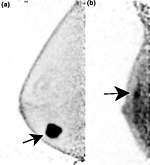
 Two PEM images, including sites of tracer uptake | |
| Purpose | imaging modality used to detect breast cancer |
PEM is not recommended for routine use or for breast cancer screening, in part due to higher radiation dose compared to other modalities.[4] Compared to breast MRI, PEM offers higher specificity.[5] Specific indications can include "high-risk patients with masses > 2 cm or aggressive malignancy and serum tumor marker elevation".[6][7] 18F-FDG is the most common radiopharmaceutical used for PEM.[8]
Equipment
.png)
PEM uses a specialised scanning system. Though some systems resemble a small PET scanner with a ring of detectors, others consist of a pair of gamma radiation detectors placed above and below the breast.[9] On these systems, mild breast compression is applied to spread the breast and reduce its thickness. The detection process is identical to standard PET scanners. Positrons emitted by the injected 18F-FDG annihilate on interaction with electrons in tissue, leading to the emission of a pair of photons travelling in opposite directions. The detection of two simultaneous photons indicates the emission of a positron at a point on the line linking the two detection events. An image is the reconstructed from the collected emission data.[10][11]
History
Mammography using positron emitters was first proposed in 1994.[12] PEM is now approved in the United States and Europe for post-diagnosis imaging, with multiple commercial systems available.[13][14]
See also
References
- MacDonald, L.; Edwards, J.; Lewellen, T.; Haseley, D.; Rogers, J.; Kinahan, P. (October 2009). "Clinical imaging characteristics of the positron emission mammography camera: PEM Flex Solo II". J. Nucl. Med. 50 (10): 1666–75. doi:10.2967/jnumed.109.064345. PMC 2873041. PMID 19759118.
- Specht, Jennifer M; Mankoff, David A (16 March 2012). "Advances in molecular imaging for breast cancer detection and characterization". Breast Cancer Research. 14 (2): 206. doi:10.1186/bcr3094. PMC 3446362. PMID 22423895.
- Marino, Maria Adele; Helbich, Thomas H.; Blandino, Alfredo; Pinker, Katja (9 June 2015). "The role of positron emission tomography in breast cancer: a short review". Memo - Magazine of European Medical Oncology. 8 (2): 130–135. doi:10.1007/s12254-015-0210-z.
- Drukteinis, Jennifer S.; Mooney, Blaise P.; Flowers, Chris I.; Gatenby, Robert A. (June 2013). "Beyond Mammography: New Frontiers in Breast Cancer Screening". The American Journal of Medicine. 126 (6): 472–479. doi:10.1016/j.amjmed.2012.11.025. PMC 4010151. PMID 23561631.
- Kalles, Vasileios; Zografos, George C.; Provatopoulou, Xeni; Koulocheri, Dimitra; Gounaris, Antonia (13 December 2012). "The current status of positron emission mammography in breast cancer diagnosis". Breast Cancer. 20 (2): 123–130. doi:10.1007/s12282-012-0433-3. PMID 23239242.
- Fletcher, J. W.; Djulbegovic, B.; Soares, H. P.; Siegel, B. A.; Lowe, V. J.; Lyman, G. H.; Coleman, R. E.; Wahl, R.; Paschold, J. C.; Avril, N.; Einhorn, L. H.; Suh, W. W.; Samson, D.; Delbeke, D.; Gorman, M.; Shields, A. F. (20 February 2008). "Recommendations on the Use of 18F-FDG PET in Oncology". Journal of Nuclear Medicine. 49 (3): 480–508. doi:10.2967/jnumed.107.047787. PMID 18287273.
- Hosono, Makoto; Saga, Tsuneo; Ito, Kengo; Kumita, Shinichiro; Sasaki, Masayuki; Senda, Michio; Hatazawa, Jun; Watanabe, Hiroshi; Ito, Hiroshi; Kanaya, Shinichi; Kimura, Yuichi; Saji, Hideo; Jinnouchi, Seishi; Fukukita, Hiroyoshi; Murakami, Koji; Kinuya, Seigo; Yamazaki, Junichi; Uchiyama, Mayuki; Uno, Koichi; Kato, Katsuhiko; Kawano, Tsuyoshi; Kubota, Kazuo; Togawa, Takashi; Honda, Norinari; Maruno, Hirotaka; Yoshimura, Mana; Kawamoto, Masami; Ozawa, Yukihiko (31 May 2014). "Clinical practice guideline for dedicated breast PET". Annals of Nuclear Medicine. 28 (6): 597–602. doi:10.1007/s12149-014-0857-2. PMC 4328123. PMID 24878887.
- Cintolo, Jessica Anna; Tchou, Julia; Pryma, Daniel A. (16 March 2013). "Diagnostic and prognostic application of positron emission tomography in breast imaging: emerging uses and the role of PET in monitoring treatment response". Breast Cancer Research and Treatment. 138 (2): 331–346. doi:10.1007/s10549-013-2451-z. PMID 23504108.
- Martins, Mónica Vieira (2015). "Positron Emission Mammography". In Fernandes, Fabiano Cavalcanti (ed.). Mammography Techniques and Review. Intech. doi:10.5772/60452. ISBN 978-953-51-2138-1.
- Glass and Shah (2013). "Clinical utility of positron emission mammography". Proc (Bayl Univ Med Cent). 26 (3): 314–9. doi:10.1080/08998280.2013.11928996. PMC 3684309. PMID 23814402.
- MacDonald, L.; Edwards, J.; Lewellen, T.; Haseley, D.; Rogers, J.; Kinahan, P. (16 September 2009). "Clinical Imaging Characteristics of the Positron Emission Mammography Camera: PEM Flex Solo II". Journal of Nuclear Medicine. 50 (10): 1666–1675. doi:10.2967/jnumed.109.064345. PMC 2873041. PMID 19759118.
- Thompson, C. J.; Murthy, K.; Weinberg, I. N.; Mako, F. (April 1994). "Feasibility study for positron emission mammography". Medical Physics. 21 (4): 529–538. doi:10.1118/1.597169. PMID 8058019.
- "Pros and Cons of Molecular Breast Imaging Tools". Imaging Technology News. 25 July 2012. Retrieved 5 January 2018.
- Subramaniam, Rathan (2015). PET/CT and Patient Outcomes, Part I, An Issue of PET Clinics. Elsevier Health Sciences. p. 161. ISBN 9780323370066.
![]()