Polarized light microscopy
Polarized light microscopy can mean any of a number of optical microscopy techniques involving polarized light. Simple techniques include illumination of the sample with polarized light. Directly transmitted light can, optionally, be blocked with a polariser orientated at 90 degrees to the illumination. More complex microscopy techniques which take advantage of polarized light include differential interference contrast microscopy and interference reflection microscopy. Scientists will often use a device called a polarizing plate to convert natural light into polarized light.[1]
These illumination techniques are most commonly used on birefringent samples where the polarized light interacts strongly with the sample and so generating contrast with the background. Polarized light microscopy is used extensively in optical mineralogy.
The Michel-Levy chart
.jpg)
As polarised light passes through a birefringent sample, the phase difference between the fast and slow directions varies with the thickness, and wavelength of light used. The optical path difference (o.p.d.) is defined as , where t is the thickness of the sample.

This then leads to a phase difference between the light passing in the two vibration directions of . For example, if the optical path difference is , then the phase difference will be , and so the polarisation will be perpendicular to the original, resulting in all of the light passing through the analyser for crossed polars. If the optical path difference is , then the phase difference will be , and so the polarisation will be parallel to the original. This means that no light will be able to pass through the analyser which it is now perpendicular to.


The Michel-Levy Chart (named after Auguste Michel-Lévy) arises when polarised white light is passed through a birefringent sample. If the sample is of uniform thickness, then only one specific wavelength will meet the above condition described above, and be perpendicular to the direction of the analyser. This means that instead of polychromatic light being viewed at the analyser, one specific wavelength will have been removed. This information can be used in a number of ways:
- If the birefringence is known, then the thickness, t, of the sample can be determined
- If the thickness is known, then the birefringence of the sample can be determined
As the order of the optical path difference increases, then it is more likely that more wavelengths of light will be removed from the spectrum. This results in the appearance of the colour being "washed out", and it becomes more difficult to determine the properties of the sample. This, however, only occurs when the sample is relatively thick when compared to the wavelength of light.
- Comparison of transilumination techniques used to generate contrast in a sample of tissue paper. 1.559 μm/pixel.
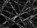 Cross-polarized light illumination, sample contrast comes from rotation of polarized light through the sample.
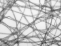 Bright field illumination, sample contrast comes from absorbance of light in the sample.
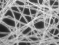 Dark field illumination, sample contrast comes from light scattered by the sample.
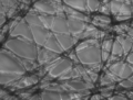 Phase contrast illumination, sample contrast comes from interference of different path lengths of light through the sample.
See also
- Petrographic microscope
References
- "Basics of Polarizing Microscopy" (PDF). Olympus. Archived (PDF) from the original on 15 December 2016. Retrieved 15 December 2016.
External links
- Polarized light microscope (Université Paris Sud)