Pneumatocele
A pneumatocele is a cavity in the lung parenchyma filled with air that may result from pulmonary trauma during mechanical ventilation.[1]
Gas-filled, or air-filled lesions in bone are known as pneumocysts.[2] When a pneumocyst is found in a bone it is called an intraosseous pneumocyst, or a vertebral pneumocyst when found in a vertebra.[3]
Cause
A pneumatocele results when a lung laceration, a cut or tear in the lung tissue, fills with air.[4] A rupture of a small airway creates the air-filled cavity.[1] Pulmonary lacerations that fill with blood are called pulmonary hematomas.[4] In some cases, both pneumatoceles and hematomas exist in the same injured lung.[5] A pneumatocele can become enlarged, for example when the patient is mechanically ventilated or has acute respiratory distress syndrome, in which case it may not go away for months.[5]
Intraosseous pneumatocysts in the bone are rare and of unclear origin. They are benign and usually without symptoms.[3] They are also found around a sacroiliac joint, and there has been one reported case of an acetabular pneumocyst.[6]
Diagnosis
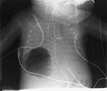
Diagnosis can be made using chest X-ray; the lesion shows up as a small, round area filled with air.[1] Computed tomography can give a more detailed understanding of the lesion.[1] Differential diagnoses – other conditions that could cause similar symptoms as pneumatocele include lung cancer, tuberculosis[7], and a lung abscess[1] in the setting of hyper IgE syndrome (aka Job's syndrome) or on its own, often caused by Staphylococcus aureus infection during cystic fibrosis.
Management and treatment
Treatment typically is supportive and includes monitoring and observation.[1]
References
- Atluri P, Karakousis GC, Porrett PM, Kaiser LR (2005). The Surgical Review: An Integrated Basic and Clinical Science Study Guide (Recall Series). Hagerstown, MD: Lippincott Williams & Wilkins. p. 376. ISBN 0-7817-5641-3.
- "pneumatocyst | Definition of pneumatocyst in English by Oxford Dictionaries". Oxford Dictionaries | English. Retrieved 4 June 2019.
- Al-Tarawneh, E; Al-Qudah, M; Hadidi, F; Jubouri, S; Hadidy, A (March 2014). "Incidental intraosseous pneumatocyst with gas-density-fluid level in an adolescent: a case report and review of the literature". Journal of Radiology Case Reports. 8 (3): 16–22. doi:10.3941/jrcr.v8i3.1540. PMC 4035364. PMID 24967024.
- White C, Stern EJ (1999). Chest Radiology Companion. Hagerstown, MD: Lippincott Williams & Wilkins. pp. 80, 176. ISBN 0-397-51732-7. Retrieved 2008-04-30.
- Gavelli G, Canini R, Bertaccini P, Battista G, Bnà C, Fattori R (June 2002). "Traumatic injuries: imaging of thoracic injuries". European Radiology. 12 (6): 1273–1294. doi:10.1007/s00330-002-1439-6. PMID 12042932.
- Narváez, JA; Narváez, J; Rodríguez-Mijarro, M; Quintero, JC (1999). "Acetabular pneumatocyst containing air-fluid level". European Radiology. 9 (8): 1647–9. doi:10.1007/s003300050902. PMID 10525883.
- Puri, M. M.; Jain, A. K.; Kumar, Lokender; Sarin, R. (April 2014). "Total replacement of a lung by tuberculosis pneumatocele--an unusual post-tuberculosis sequel". The Indian Journal of Tuberculosis. 61 (2): 162–165. ISSN 0019-5707. PMID 25509941.
Further reading
Al-Tarawneh, Emad; AL-Qudah, Mohammad; Hadidi, Fadi (March 2014). "Incidental Intraosseous Pneumatocyst with gas-density-fluid level in an adolescent: a case report and review of the literature". Journal of Radiology Case Reports. 8 (3): 16–22. doi:10.3941/jrcr.v8i3.1540. PMC 4035364. PMID 24967024.