Plasmodium falciparum
Plasmodium falciparum is a unicellular protozoan parasite of humans, and the deadliest species of Plasmodium that causes malaria in humans.[2] The parasite is transmitted through the bite of a female Anopheles mosquito and causes the disease's most dangerous form called falciparum malaria which is responsible for around 50% of all malaria cases.[3][4] P. falciparum is therefore regarded as the deadliest parasite in humans, causing 435,000 deaths in 2017.[5] It is also associated with the development of blood cancer (Burkitt's lymphoma) and is classified as Group 2A carcinogen.
| Plasmodium falciparum | |
|---|---|
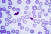 | |
| Macrogametocyte (left) and microgametocyte (right) of P. falciparum | |
| Scientific classification | |
| (unranked): | Diaphoretickes |
| Clade: | TSAR |
| Clade: | SAR |
| Infrakingdom: | Alveolata |
| Phylum: | Apicomplexa |
| Class: | Aconoidasida |
| Order: | Haemospororida |
| Family: | Plasmodiidae |
| Genus: | Plasmodium |
| Species: | P. falciparum |
| Binomial name | |
| Plasmodium falciparum Welch, 1897 | |
| Synonyms[1] | |
| |
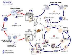
The species originated from the malarial parasite Laverania found in gorillas, around 10,000 years ago.[6] Alphonse Laveran was the first to identify the parasite in 1880, and named it Oscillaria malariae. Ronald Ross discovered its transmission by mosquito in 1897. Giovanni Battista Grassi elucidated the complete transmission from a female anopheline mosquito to humans in 1898. In 1897, William H. Welch created the name Plasmodium falciparum, which ICZN formally adopted in 1954. P. falciparum assumes several different forms during its life cycle. The human-infective stage are sporozoites from the salivary gland of a mosquito. The sporozoites grow and multiply in the liver to become merozoites. These merozoites invade the erythrocytes (RBCs) to form trophozoites, schizonts and gametocytes, during which the symptoms of malaria are produced. In the mosquito, the gametocytes undergo sexual reproduction to a zygote, which turns into ookinete. Ookinete forms oocyts from which sporozoites are formed.
As of the latest World Malaria Report of the World Health Organization, there were 219 million cases of malaria worldwide in 2017, up from 216 million cases in 2016. This resulted in an estimated 435,000 deaths.[5] Almost every malarial death is caused by P. falciparum, and 93% of death occurs in Africa. Children under five years of age are most affected, accounting for 61% of the total deaths.[5] In Sub-Saharan Africa, over 75% of cases were due to P. falciparum, whereas in most other malarial countries, other, less virulent plasmodial species predominate.[7]
History
Falciparum malaria was familiar to the ancient Greeks, who gave the general name πυρετός pyretós "fever".[8] Hippocrates (c. 460–370 BCE) gave several descriptions on tertian fever and quartan fever.[9] It was prevalent throughout the ancient Egyptian and Roman civilizations.[10] It was the Romans who named the disease "malaria"—mala for bad, and aria for air, as they believed that the disease was spread by contaminated air, or miasma.
Discovery
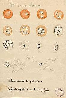
A German physician, Johann Friedrich Meckel, must have been the first to see P. falciparum but without knowing what it was. In 1847 he reported the presence of black pigment granules from the blood and spleen of a patient who died of malaria. The French Army physician Charles Louis Alphonse Laveran, while working at Bône Hospital (now Annaba in Algeria), correctly identified the parasite as a causative pathogen of malaria in 1880. He presented his discovery before the French Academy of Medicine in Paris, and published it in The Lancet, in 1881. He gave the scientific name Oscillaria malariae.[11] But his discovery was received with skepticism mainly because by that time leading physicians such as Theodor Albrecht Edwin Klebs and Corrado Tommasi-Crudeli claimed that they had discovered a bacterium (which they called Bacillus malariae) as the pathogen of malaria. Laveran's discovery was widely accepted only after five years when Camillo Golgi confirmed the parasite using better microscope and staining technique. Laveran was awarded the Nobel Prize in Physiology or Medicine in 1907 for his work. In 1900, the Italian zoologist Giovanni Battista Grassi categorized Plasmodium species based on the timing of fever in the patient; malignant tertian malaria was caused by Laverania malariae (now P. falciparum), benign tertian malaria by Haemamoeba vivax (now P. vivax), and quartan malaria by Haemamoeba malariae (now P. malariae).[12]
The British physician Patrick Manson formulated the mosquito-malaria theory in 1894; until that time, malarial parasites were believed to be spread in air as miasma, a Greek word for pollution.[11] His colleague Ronald Ross, a British Army surgeon, travelled to India to test the theory. Ross discovered in 1897 that malarial parasites lived in certain mosquitoes. The next year, he demonstrated that a malarial parasite of birds could be transmitted by mosquitoes from one bird to another. Around the same time, Grassi demonstrated that P. falciparum was transmitted in humans only by female anopheline mosquito (in his case Anopheles claviger).[13] Ross, Manson and Grassi were nominated for the Nobel Prize in Physiology or Medicine in 1902. Under controversial circumstances, only Ronald Ross was selected for the award.[14]
There was a long debate on the taxonomy. It was only in 1954 the International Commission on Zoological Nomenclature officially approved the binominal Plasmodium falciparum.[15] The valid genus Plasmodium was created by two Italian physicians Ettore Marchiafava and Angelo Celli in 1885. The species name was introduced by an American physician William Henry Welch in 1897.[16] It is derived from the Latin falx, meaning "sickle" and parum meaning "like or equal to another".[15]
Origin and evolution
P. falciparum is now generally accepted to have evolved from Laverania (a subgenus of Plasmodium found in apes) species present in gorilla in Western Africa.[17][18] Genetic diversity indicates that the human protozoan emerged around 10,000 years ago.[6] The closest relative of P. falciparum is P. praefalciparum, a parasite of gorillas, as supported by mitochondrial, apicoplastic and nuclear DNA sequences.[19][20][21] These two species are closely related to the chimpanzee parasite P. reichenowi, which was previously thought to be the closest relative of P. falciparum. P. falciparum was also once thought to originate from a parasite of birds.[22]
Levels of genetic polymorphism are extremely low within the P. falciparum genome compared to that of closely related, ape infecting species of Plasmodium (including P. praefalciparum)[23][19]. This suggests that the origin of P. falciparum in humans is recent, as a single P. praefalciparum strain became capable of infecting humans.[19] The genetic information of Plasmodium falciparum has signaled a recent expansion that coincides with the agricultural revolution. It is likely that the development of extensive agriculture increased mosquito population densities by giving rise to more breeding sites, which may have triggered the evolution and expansion of Plasmodium falciparum.[24]
Structure
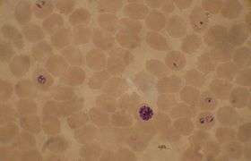
P. falciparum does not have a fixed structure but undergoes continuous change during the course of its life cycle. A sporozoite is spindle-shaped and 10-15 μm long. In the liver it grows into an ovoid schizont of 30-70 μm in diameter. Each schizont produces merozoites, each of which is roughly 1.5 μm in length and 1 μm in diameter. In the erythrocyte the merozoite form a ring-like structure, becoming a trophozoite. A trophozoites feed on the haemoglobin and forms a granular pigment called haemozoin. Unlike those of other Plasmodium species, the gametocytes of P. falciparum are elongated and crescent-shaped, by which they are sometimes identified. A mature gametocyte is 8-12 μm long and 3-6 μm wide. The ookinete is also elongated measuring about 18-24 μm. An oocyst is rounded and can grow up to 80 μm in diameter.[25] Microscopic examination of a blood film reveals only early (ring-form) trophozoites and gametocytes that are in the peripheral blood. Mature trophozoites or schizonts in peripheral blood smears, as these are usually sequestered in the tissues. On occasion, faint, comma-shaped, red dots are seen on the erythrocyte surface. These dots are Maurer's cleft and are secretory organelles that produce proteins and enzymes essential for nutrient uptake and immune evasion processes.[26]
The apical complex, which is actually a combination of organelles, is an important structure. It contains secretory organelles called rhoptries and micronemes, which are vital for mobility, adhesion, host cell invasion, and parasitophorous vacuole formation.[27] As an apicomplexan, it harbours a plastid, an apicoplast, similar to plant chloroplasts, which they probably acquired by engulfing (or being invaded by) a eukaryotic alga and retaining the algal plastid as a distinctive organelle encased within four membranes. The apicoplast is involved in the synthesis of lipids and several other compounds and provides an attractive drug target. During the asexual blood stage of infection, an essential function of the apicoplast is to produce the isoprenoid precursors isopentenyl pyrophosphate (IPP) and dimethylallyl pyrophosphate (DMAPP) via the MEP (non-mevalonate) pathway .[28]
Genome
In 1995 the Malaria Genome Project was set up to sequence the genome of P. falciparum. The genome of its mitochondrion was reported in 1995, that of the nonphotosynthetic plastid known as the apicoplast in 1996,[29] and the sequence of the first nuclear chromosome (chromosome 2) in 1998. The sequence of chromosome 3 was reported in 1999 and the entire genome was reported on 3 October 2002.[30] The roughly 24-megabase genome is extremely AT-rich (about 80%) and is organised into 14 chromosomes. Just over 5,300 genes were described. Many genes involved in antigenic variation are located in the subtelomeric regions of the chromosomes. These are divided into the var, rif, and stevor families. Within the genome, there exist 59 var, 149 rif, and 28 stevor genes, along with multiple pseudogenes and truncations. It is estimated that 551, or roughly 10%, of the predicted nuclear-encoded proteins are targeted to the apicoplast, while 4.7% of the proteome is targeted to the mitochondria.[30]
Life cycle
Humans are the intermediate hosts in which asexual reproduction occurs, and female anopheline mosquitos are the definitive hosts harbouring the sexual reproduction stage.
In humans
Infection in humans begins with the bite of an infected female Anopheles mosquito. Out of about 460 species of Anopheles mosquito, more than 70 species transmit falciparum malaria.[31] Anopheles gambiae is one of the best known and most prevalent vectors, particularly in Africa.[32]
The infective stage called sporozoites released from the salivary glands through the proboscis of the mosquito enter the bloodstream during feeding. The mosquito saliva contains antihemostatic and anti-inflammatory enzymes that disrupt blood clotting and inhibit the pain reaction. Typically, each infected bite contains 20-200 sporozoites.[27] The immune system clears the sporozoites from the circulation within 30 minutes. But a few escape and quickly invade liver cells (hepatocytes).[33] The sporozoites move in the blood stream by gliding, which is driven by motor made up of proteins actin and myosin beneath their plasma membrane.[34]
Liver stage or exo-erythrocytic schizogony
Entering the hepatocytes, the parasite loses its apical complex and surface coat, and transforms into a trophozoite. Within the parasitophorous vacuole of the hepatocyte, it undergoes 13-14 rounds of mitosis and meiosis which produce a syncytial cell (coenocyte) called a schizont. This process is called schizogony. A schizont contains tens of thousands of nuclei. From the surface of the schizont, tens of thousands of haploid (1n) daughter cells called merozoites emerge. The liver stage can produce up to 90,000 merozoites,[35] which are eventually released into the bloodstream in parasite-filled vesicles called merosomes.[36]
Blood stage or erythrocytic schizogony
Merozoites use the apicomplexan invasion organelles (apical complex, pellicle and surface coat) to recognize and enter the host erythrocyte (red blood cell). The parasite first binds to the erythrocyte in a random orientation. It then reorients such that the apical complex is in proximity to the erythrocyte membrane. The parasite forms a parasitophorous vacuole, to allow for its development inside the erythrocyte.[37] This infection cycle occurs in a highly synchronous fashion, with roughly all of the parasites throughout the blood in the same stage of development. This precise clocking mechanism has been shown to be dependent on the human host's own circadian rhythm.[38]
Within the erythrocyte, the parasite metabolism depends on the digestion of hemoglobin. The clinical symptoms of malaria such as fever, anemia, and neurological disorder are produced during the blood stage.[33]
The parasite can also alter the morphology of the erythrocyte, causing knobs on the erythrocyte membrane. Infected erythrocytes are often sequestered in various human tissues or organs, such as the heart, liver and brain. This is caused by parasite-derived cell surface proteins being present on the erythrocyte membrane, and it is these proteins that bind to receptors on human cells. Sequestration in the brain causes cerebral malaria, a very severe form of the disease, which increases the victim's likelihood of death.
Trophozoite
After invading the erythrocyte, the parasite loses its specific invasion organelles (apical complex and surface coat) and de-differentiates into a round trophozoite located within a parasitophorous vacuole. The young trophozoite (or "ring" stage, because of its morphology on stained blood films) grows substantially before undergoing schizogony.[39]
Schizont
At the schizont stage, the parasite replicates its DNA multiple times and multiple mitotic divisions occur asynchronously.[40][41] Each schizont forms 16-18 merozoites.[39] The red blood cells are ruptured by the merozoites. The liberated merozoites invade fresh erythrocytes. A free merozoite is in the bloodstream for roughly 60 seconds before it enters another erythrocyte.[37]
The duration of each blood stage is approximately 48 hours. This gives rise to the characteristic clinical manifestations of falciparum malaria, such as fever and chills, corresponding to the synchronous rupture of the infected erythrocytes.[42]
Gametocyte
Not all of the merozoites divide into schizonts; some get differentiated into sexual forms, male and female gametocytes. These gametocytes take roughly 7–15 days to reach full maturity, through the process called gametocytogenesis. These gametocytes are taken up by a female Anopheles mosquito during a blood meal.[43]
Incubation period
The time of appearance of the symptoms from infection (called incubation period) is shortest for P. falciparum among Plasmodium species. An average incubation period is 11 days,[42] but may range from 9 to 30 days. In isolated cases, prolonged incubation period as long as 2, 3 or even 8 years have been recorded.[44] Pregnancy and co-infection with HIV are important conditions for delayed symptoms.[45] Parasites can be detected from blood samples by the 10th day after infection (pre-patent period).[42]
In mosquitoes
Within the mosquito midgut, the female gamete maturation process entails slight morphological changes, becoming more enlarged and spherical. The male gametocyte undergoes a rapid nuclear division within 15 minutes, producing eight flagellated microgametes by a process called exflagellation.[46] The flagellated microgamete fertilizes the female macrogamete to produce a diploid cell called a zygote. The zygote then develops into an ookinete. The ookinete is a motile cell, capable of invading other organs of the mosquito. It traverses the peritrophic membrane of the mosquito midgut and crosses the midgut epithelium. Once through the epithelium, the ookinete enters the basal lamina, and settles to an immotile oocyst. For several days, the oocyst undergoes 10 to 11 rounds of cell division to create a syncytial cell (sporoblast) containing thousands of nuclei. Meiosis takes place inside the sporoblast to produce over 3,000 haploid daughter cells called sporozoites on the surface of the mother cell.[47] Immature sporozoites break through the oocyst wall into the haemolymph. They migrate to the mosquito salivary glands where they undergo further development and become infective to humans.[33]
Interaction with human immune system
Immune response
A single anopheline mosquito can transmit hundreds of P. falciparum sporozoites in a single bite under experimental conditions. But in nature the number is generally less than 80.[48] The sporozoites do not enter the blood stream directly and remain in the skin tissue for 2 to 3 hours. About 15–20% sporozoites enter the lymphatic system where they activate dendritic cells, which send them for destruction by T lymphocytes (CD8+ T cells). At 48 hours after infection, Plasmodium-specific CD8+ T cells can be detected in the lymph nodes connected to the skin cells.[49] Most of the sporozites remaining in the skin tissue are subsequently killed by the innate immune system. The sporozoite glycoprotein specifically activates mast cells. The mast cells then produce signalling molecules such as TNFα and MIP-2, which activate cell eaters (professional phagocytes) such as neutrophils and macrophages.[50]
Only a small number (0.5-5%) of sporozoites enter the blood stream into the liver. In the liver, the activated CD8+ T cells from the lymph bind the sporozoites through the circumsporozoite protein (CSP).[49] Antigen presentation by dendritic cells in the skin tissue to T cells is also a crucial process. From this stage onward the parasites produce different proteins that help in suppressing communication of the immune cells.[51] Even at the height of the infection when RBCs are ruptured, the immune signals are not strong enough to activate macrophages or natural killer cells.[52]
Immune system evasion
Although P. falciparum is easily recognized by human immune system while in the bloodstream, it evades immunity by producing over 2,000 cell membrane antigens[53] The initial infective stage sporozoites produce circumsporozoite protein (CSP), which binds to hepatocytes.[54] Binding to and entry into the hepatocytes is aided by another protein, thrombospondin-related anonymous protein (TRAP).[55] TRAP and other secretory proteins (including sporozoite microneme protein essential for cell traversal 1, SPECT1 and SPECT2) from microneme allow the sporozoite to move through the blood, avoiding immune cells and penetrating hepatocytes.[56]
During erythrocyte invasion, merozoites release merozoite cap protein-1 (MCP1), apical membrane antigen 1 (AMA1), erythrocyte-binding antigens (EBA), myosin A tail domain interacting protein (MTIP), and merozoite surface proteins (MSPs).[53] Of these MSPs, MSP1 and MSP2 are primarily responsible for avoiding immune cells.[57] The virulence of P. falciparum is mediated by erythrocyte membrane proteins, which are produced by the schizonts and trophozoites inside the erythrocytes and are displayed on the erythrocyte membrane. PfEMP1 is the most important, capable of acting as both an antigen and an adhesion molecule.[58]
Pathogenesis
The clinical symptoms of falciparum malaria are produced by the rupture of schizont and destruction of erythrocytes. Most of the patients experience fever (>92% of cases), chills (79%), headaches (70%), and sweating (64%). Dizziness, malaise, muscle pain, abdominal pain, nausea, vomiting, mild diarrhea, and dry cough are also generally associated. High heartrate, jaundice, pallor, orthostatic hypotension, enlarged liver, and enlarged spleen are also diagnosed.[42]
P. falciparum works via sequestration, a distinctive property not shared by few other Plasmodiae. The mature schizonts change the surface properties of infected erythrocytes, causing them to stick to blood vessel walls (cytoadherence). This leads to obstruction of the microcirculation and results in dysfunction of multiple organs, such as the brain in cerebral malaria.[59]
P. falciparum is responsible for (almost) all severe human illnesses and deaths due to malaria, in a condition called complicated or severe malaria. Complicated malaria occurs more commonly in children under age 5,[42] and sometimes in pregnant women (a condition specifically called pregnancy-associated malaria).[60] Women become susceptible to severe malaria during their first pregnancy. Susceptibility to severe malaria is reduced in subsequent pregnancies due to increased antibody levels against variant surface antigens that appear on infected erythrocytes.[61] But increased immunity in mother increases susceptibility to malaria in newborn babies.[60]
Distribution and epidemiology
P. falciparum is found in all continents except Europe. According to the WHO World Malaria Report 2018, 219 million people suffered from malaria in 2017, an increase from 216 million in 2016. 435,000 people died from it. The infection is most prevalent in Africa, where 92% of malaria deaths occur. Children under five years of age are most affected and 61% of malaria deaths occurred in this age group. 80% of the infection is found in Sub-Saharan Africa, 7% in the South-East Asia, and 2% in the Eastern Mediterranean. Nigeria has the highest incidence with 27% of the total global cases. Outside Africa, India has the highest incidence with 4.5% of the global burden.[62] Europe is regarded as a malaria-free region. Historically, the parasite and its disease had been most well known in Europe. But medical programmes, such as insecticide spraying, drug therapy and environmental engineering since the early 20th century resulted in complete eradication in the 1970s.[63] It is estimated that approximately 2.4 billion people are at constant risk of infection.[64]
Treatment
History
In 1640, Huan del Vego first employed the tincture of the cinchona bark for treating malaria; the native Indians of Peru and Ecuador had been using it even earlier for treating fevers. Thompson (1650) introduced this "Jesuits' bark" to England. Its first recorded use there was by John Metford of Northampton in 1656. Morton (1696) presented the first detailed description of the clinical picture of malaria and of its treatment with cinchona. Gize (1816) studied the extraction of crystalline quinine from the cinchona bark and Pelletier and Caventou (1820) in France extracted pure quinine alkaloids, which they named quinine and cinchonine.[65][66] The total synthesis of quinine was achieved by American chemists R.B. Woodward and W.E. Doering in 1944. Woodward received the Nobel Prize in Chemistry in 1965.[67]
Attempts to make synthetic antimalarials began in 1891. Atabrine, developed in 1933, was used widely throughout the Pacific in World War II, but was unpopular because of its adverse effects.[68] In the late 1930s, the Germans developed chloroquine, which went into use in the North African campaigns. Creating a secret military project called Project 523, Mao Zedong encouraged Chinese scientists to find new antimalarials after seeing the casualties in the Vietnam War. Tu Youyou discovered artemisinin in the 1970s from sweet wormwood (Artemisia annua). This drug became known to Western scientists in the late 1980s and early 1990s and is now a standard treatment. Tu won the Nobel Prize in Physiology or Medicine in 2015.[69]
Uncomplicated malaria
According to WHO guidelines 2010,[70] artemisinin-based combination therapies (ACTs) are the recommended first-line antimalarial treatments for uncomplicated malaria caused by P. falciparum. WHO recommends combinations such as artemether/lumefantrine, artesunate/amodiaquine, artesunate/mefloquine, artesunate/sulfadoxine-pyrimethamine, and dihydroartemisinin/piperaquine.[70]
The choice of ACT is based on the level of resistance to the constituents in the combination. Artemisinin and its derivatives are not appropriate for monotherapy. As second-line antimalarial treatment, when initial treatment does not work, an alternative ACT known to be effective in the region is recommended, such as artesunate plus tetracycline or doxycycline or clindamycin, and quinine plus tetracycline or doxycycline or clindamycin. Any of these combinations is to be given for 7 days. For pregnant women, the recommended first-line treatment during the first trimester is quinine plus clindamycin for 7 days.[70] Artesunate plus clindamycin for 7 days is indicated if this treatment fails. For travellers returning to nonendemic countries, atovaquone/proguanil, artemether/lumefantrineany and quinine plus doxycycline or clindamycin are recommended.[70]
Severe malaria
For adults, intravenous (IV) or intramuscular (IM) artesunate is recommended.[70] Quinine is an acceptable alternative if parenteral artesunate is not available.[70]
For children, especially in the malaria-endemic areas of Africa, artesunate IV or IM, quinine (IV infusion or divided IM injection), and artemether IM are recommended.[70]
Parenteral antimalarials should be administered for a minimum of 24 hours, irrespective of the patient's ability to tolerate oral medication earlier.[70] Thereafter, complete treatment is recommended including complete course of ACT or quinine plus clindamycin or doxycycline.[70]
Vaccination
RTS,S is the only candidate as malaria vaccine to have gone through clinical trials.[71] Analysis of the results of the phase III trial (conducted between 2011 and 2016) revealed a rather low efficacy (20-39% depending on age, with up to 50% in 5–17-month aged babies), indicating that the vaccine will not lead to full protection and eradication.[72]
Cancer
The International Agency for Research on Cancer (IARC) has classified malaria due to P. falciparum as Group 2A carcinogen, meaning that the parasite is probably a cancer-causing agent in humans.[73] Its association with a blood cell (lymphocyte) cancer called Burkitt's lymphoma is established. Burkit's lymphoma was discovered by Denis Burkitt in 1958 from African children, and he later speculated that the cancer was likely due to certain infectious diseases. In 1964, a virus, later called Epstein–Barr virus (EBV) after the discoverers, was identified from the cancer cells. The virus was subsequently proved to be the direct cancer agent, and is now classified as Group 1 carcinogen.[74] In 1989, it was realised that EBV requires other infections such as with malaria to cause lymphocyte transformation. It was reported that the incidence of Burkitt's lymphoma decreased with effective treatment of malaria over several years.[75] The actual role played by P. falciparum remained unclear for the next two-and-half decades. EBV had been known to induce lymphocytes to become cancerous using its viral proteins (antigens such as EBNA-1, EBNA-2, LMP-1, and LMP2A).[76][77] From 2014, it became clear that P. falciparum contributes to the development of the lymphoma. P. falciparum-infected erythrocytes directly bind to B lymphocytes through the CIDR1α domain of PfEMP1. This binding activates toll-like receptors (TLR7 and TLR10) causing continuous activation of lymphocytes to undergo proliferation and differentiation into plasma cells, thereby increasing the secretion of IgM and cytokines.[78] This in turn activates an enzyme called activation-induced cytidine deaminase (AID), which tends to cause mutation in the DNA (by double-strand break) of an EBV-infected lymphocytes. The damaged DNA undergoes uncontrolled replication, thus making the cell cancerous.[79]
Influence on the human genome
The high mortality and morbidity caused by P. falciparum has placed great selective pressure on the human genome. Several genetic factors provide some resistance to Plasmodium infection, including sickle cell trait, thalassaemia traits, glucose-6-phosphate dehydrogenase deficiency, and the absence of Duffy antigens on red blood cells.[80][81] E. A. Beet, a doctor working in Southern Rhodesia (now Zimbabwe) had observed in 1948 that sickle-cell disease was related to lower rate of malaria infection.[82] This suggestion was reiterated by J. B. S. Haldane in 1948, who suggested that thalassaemia could provide similar protection.[83] This hypothesis has since been confirmed and extended to hemoglobin E,[84] hemoglobin C and Hemoglobin S.[85]
See also
- Malaria Atlas Project
- List of parasites (human)
- UCSC Malaria Genome Browser
References
- Coatney GR, Collins WE, Warren M, Contacos PG (1971). "22 Plasmodium falciparum (Welch, 1897)". The primate malarias. Division of Parasitic Disease, CDC. p. 263.
- Rich, S. M.; Leendertz, F. H.; Xu, G.; Lebreton, M.; Djoko, C. F.; Aminake, M. N.; Takang, E. E.; Diffo, J. L. D.; Pike, B. L.; Rosenthal, B. M.; Formenty, P.; Boesch, C.; Ayala, F. J.; Wolfe, N. D. (2009). "The origin of malignant malaria". Proceedings of the National Academy of Sciences. 106 (35): 14902–14907. Bibcode:2009PNAS..10614902R. doi:10.1073/pnas.0907740106. PMC 2720412. PMID 19666593.
- Perkins, D. J.; Were, T.; Davenport, G. C.; Kempaiah, P.; Hittner, J. B.; Ong'Echa, J. M. (2011). "Severe malarial anemia: Innate immunity and pathogenesis". International Journal of Biological Sciences. 7 (9): 1427–1442. doi:10.7150/ijbs.7.1427. PMC 3221949. PMID 22110393.
- Perlmann, P; Troye-Blomberg, M (2000). "Malaria blood-stage infection and its control by the immune system". Folia Biologica. 46 (6): 210–8. PMID 11140853.
- "World malaria report 2018". WHO. Retrieved 2 December 2018.
- Loy, Dorothy E.; Liu, Weimin; Li, Yingying; Learn, Gerald H.; Plenderleith, Lindsey J.; Sundararaman, Sesh A.; Sharp, Paul M.; Hahn, Beatrice H. (2017). "Out of Africa: origins and evolution of the human malaria parasites Plasmodium falciparum and Plasmodium vivax". International Journal for Parasitology. 47 (2–3): 87–97. doi:10.1016/j.ijpara.2016.05.008. PMC 5205579. PMID 27381764.
- "World Malaria Report 2008" (PDF). World Health Organisation. 2008. p. 10. Retrieved 2009-08-17.
- Baron, Christopher; Hamlin, Christopher (2015). "Malaria and the Decline of Ancient Greece: Revisiting the Jones Hypothesis in an Era of Interdisciplinarity". Minerva. 53 (4): 327–358. doi:10.1007/s11024-015-9280-7.
- Hempelmann, Ernst; Krafts, Kristine (2013). "Bad air, amulets and mosquitoes: 2,000?years of changing perspectives on malaria". Malaria Journal. 12 (1): 232. doi:10.1186/1475-2875-12-232. PMC 3723432. PMID 23835014.
- Nerlich, A (2016). Paleopathology and Paleomicrobiology of Malaria. Microbiology Spectrum. 4. pp. 155–160. doi:10.1128/microbiolspec.PoH-0006-2015. ISBN 9781555819163. PMID 27837743.
- Lalchhandama, K. (2014). "The making of modern malariology: from miasma to mosquito- malaria theory" (PDF). Science Vision. 14 (1): 3–17. Archived from the original (PDF) on 2014-04-27.
- Cox, Francis EG (2010). "History of the discovery of the malaria parasites and their vectors". Parasites & Vectors. 3 (1): 5. doi:10.1186/1756-3305-3-5. PMC 2825508. PMID 20205846.

- Baccetti, B (2008). "History of the early dipteran systematics in Italy: from Lyncei to Battista Grassi". Parassitologia. 50 (3–4): 167–172. PMID 20055226.
- Capanna, E (2006). "Grassi versus Ross: who solved the riddle of malaria?". International Microbiology. 9 (1): 69–74. PMID 16636993.
- Bruce-Chwatt, L.J. (1987). "Falciparum nomenclature". Parasitology Today. 3 (8): 252. doi:10.1016/0169-4758(87)90153-0.
- Christophers, R; Sinton, JA (1938). "Correct Name of Malignant Tertian Parasite". British Medical Journal. 2 (4065): 1130–1134. doi:10.1136/bmj.2.4065.1130. PMC 2211005. PMID 20781927.
- Liu, W; Li, Y; Learn, GH; Rudicell, RS; Robertson, JD; Keele, BF; Ndjango, JB; Sanz, CM; et al. (2010). "Origin of the human malaria parasite Plasmodium falciparum in gorillas". Nature. 467 (7314): 420–5. Bibcode:2010Natur.467..420L. doi:10.1038/nature09442. PMC 2997044. PMID 20864995.
- Holmes, Edward C. (2010). "Malaria: The gorilla connection". Nature. 467 (7314): 404–405. Bibcode:2010Natur.467..404H. doi:10.1038/467404a. PMID 20864986.
- Liu, W; Y Li, GH Learn, RS Rudicell, JD Robertson, BF Keele, JN Ndjango, CM Sanz, DB Morgan, S Locatelli, MK Gonder, PJ Kranzusch, PD Walsh, E Delaporte, E Mpoudi-Ngole, AV Georgiev, MN Muller, GM Shaw, M Peeters, PM Sharp, JC Rayner, BH Hahn (2010). "Origin of the human malaria parasite Plasmodium falciparum in gorillas". Nature. 467 (7314): 420–5. Bibcode:2010Natur.467..420L. doi:10.1038/nature09442. PMC 2997044. PMID 20864995.CS1 maint: multiple names: authors list (link)
- Duval, L; M Fourment, E Nerrienet, D Rousset, SA Sadeuh, SM Goodman, NV Andriaholinirina, M Randrianarivelojosia, RE Paul, V Robert, FJ Ayala, F Ariey (2010). "African apes as reservoirs of Plasmodium falciparum and the origin and diversification of the Laverania subgenus". Proceedings of the National Academy of Sciences of the United States of America. 107 (23): 10561–6. Bibcode:2010PNAS..10710561D. doi:10.1073/pnas.1005435107. PMC 2890828. PMID 20498054.CS1 maint: multiple names: authors list (link)
- Rayner, J; WM Liu, M Peeters, PM Sharp, BH Hahn (2011). "A plethora of Plasmodium species in wild apes: a source of human infection?". Trends in Parasitology. 27 (5): 222–229. doi:10.1016/J.Pt.2011.01.006. PMC 3087880. PMID 21354860.CS1 maint: multiple names: authors list (link)
- Rathore, D; Wahl AM, Sullivan M, McCutchan TF (2001-04-25). "A phylogenetic comparison of gene trees constructed from plastid, mitochondrial and genomic DNA of Plasmodium species". Molecular and Biochemical Parasitology. 114 (1): 89–94. doi:10.1016/S0166-6851(01)00241-9. PMID 11356517.CS1 maint: multiple names: authors list (link)
- Hartl, DH (January 2004). "The origin of malaria: mixed messages from genetic diversity". Nature Reviews Microbiology. 2 (1): 15–22. doi:10.1038/nrmicro795. PMID 15035005.
- Hume, J.C.; Lyons, E.J.; Day, K.P. (2003). "Human migration, mosquitoes and the evolution of Plasmodium falciparum". Trends Parasitol. 19 (3): 144–9. doi:10.1016/s1471-4922(03)00008-4. PMID 12643998.
- Lucius, R.; Roberts, C.W. (2017). "Biology of Parasitic Protozoa". In Lucius, R.; Loos-Frank, B.; Lane, R.P.; Poulin, R.; Roberts, C.W.; Grencis, R.K. (eds.). The Biology of Parasites. John Wiley & Sons. pp. 190–198. ISBN 978-3-527-32848-2.
- Lanzer, Michael; Wickert, Hannes; Krohne, Georg; Vincensini, Laetitia; Braun Breton, Catherine (2006). "Maurer's clefts: A novel multi-functional organelle in the cytoplasm of Plasmodium falciparum-infected erythrocytes". International Journal for Parasitology. 36 (1): 23–36. doi:10.1016/j.ijpara.2005.10.001. PMID 16337634.
- Garcia, J. E.; Puentes, A.; Patarroyo, M. E. (2006). "Developmental Biology of Sporozoite-Host Interactions in Plasmodium falciparum Malaria: Implications for Vaccine Design". Clinical Microbiology Reviews. 19 (4): 686–707. doi:10.1128/CMR.00063-05. PMC 1592691. PMID 17041140.
- Yeh, Ellen; DeRisi, Joseph L. (2011-08-30). "Chemical Rescue of Malaria Parasites Lacking an Apicoplast Defines Organelle Function in Blood-Stage Plasmodium falciparum". PLOS Biol. 9 (8): e1001138. doi:10.1371/journal.pbio.1001138. ISSN 1545-7885. PMC 3166167. PMID 21912516.
- Wilson RJ; Denny PW; Preiser PR; et al. (August 1996). "Complete gene map of the plastid-like DNA of the malaria parasite Plasmodium falciparum". Journal of Molecular Biology. 261 (2): 155–72. doi:10.1006/jmbi.1996.0449. PMID 8757284.
- Gardner MJ; Hall N; Fung E; et al. (October 2002). "Genome sequence of the human malaria parasite Plasmodium falciparum". Nature. 419 (6906): 498–511. Bibcode:2002Natur.419..498G. doi:10.1038/nature01097. PMC 3836256. PMID 12368864.
- Molina-Cruz, Alvaro; Zilversmit, Martine M.; Neafsey, Daniel E.; Hartl, Daniel L.; Barillas-Mury, Carolina (2016). "Mosquito Vectors and the Globalization of Plasmodium falciparum Malaria". Annual Review of Genetics. 50 (1): 447–465. doi:10.1146/annurev-genet-120215-035211. PMID 27732796.
- Sinka, Marianne E; Bangs, Michael J; Manguin, Sylvie; Coetzee, Maureen; Mbogo, Charles M; Hemingway, Janet; Patil, Anand P; Temperley, Will H; Gething, Peter W; Kabaria, Caroline W; Okara, Robi M; Van Boeckel, Thomas; Godfray, H Charles J; Harbach, Ralph E; Hay, Simon I (2010). "The dominant Anopheles vectors of human malaria in Africa, Europe and the Middle East: occurrence data, distribution maps and bionomic pr?cis". Parasites & Vectors. 3 (1): 117. doi:10.1186/1756-3305-3-117. PMC 3016360. PMID 21129198.
- Gerald, N.; Mahajan, B.; Kumar, S. (2011). "Mitosis in the Human Malaria Parasite Plasmodium falciparum". Eukaryotic Cell. 10 (4): 474–482. doi:10.1128/EC.00314-10. PMC 3127633. PMID 21317311.
- Kappe, SH; Buscaglia, CA; Bergman, LW; Coppens, I; Nussenzweig, V (2004). "Apicomplexan gliding motility and host cell invasion: overhauling the motor model". Trends in Parasitology. 20 (1): 13–16. CiteSeerX 10.1.1.458.5746. doi:10.1016/j.pt.2003.10.011. PMID 14700584.
- Vaughan, Ashley M.; Kappe, Stefan H.I. (2017). "Malaria Parasite Liver Infection and Exoerythrocytic Biology". Cold Spring Harbor Perspectives in Medicine. 7 (6): a025486. doi:10.1101/cshperspect.a025486. PMC 5453383. PMID 28242785.
- Sturm, A. (2006). "Manipulation of Host Hepatocytes by the Malaria Parasite for Delivery into Liver Sinusoids". Science. 313 (5791): 1287–1290. Bibcode:2006Sci...313.1287S. doi:10.1126/science.1129720. PMID 16888102.
- Cowman, Alan F.; Crabb, Brendan S. (2006). "Invasion of Red Blood Cells by Malaria Parasites". Cell. 124 (4): 755–766. doi:10.1016/j.cell.2006.02.006. PMID 16497586.
- "Malaria eModule - SYNCHRONICITY".
- "Malaria eModule - ASEXUAL ERYTHROCYTIC STAGES".
- Read, M.; Sherwin, T.; Holloway, S. P.; Gull, K.; Hyde, J. E. (1993). "Microtubular organization visualized by immunofluorescence microscopy during erythrocytic schizogony in Plasmodium falciparum and investigation of post-translational modifications of parasite tubulin". Parasitology. 106 (3): 223–232. doi:10.1017/s0031182000075041.
- Arnot, D. E.; Ronander, E.; Bengtsson, D. C. (2011). "The progression of the intra-erythrocytic cell cycle of Plasmodium falciparum and the role of the centriolar plaques in asynchronous mitotic division during schizogony". Int. J. Parasitol. 41 (1): 71–80. doi:10.1016/j.ijpara.2010.07.012. PMID 20816844.
- Trampuz, Andrej; Jereb, Matjaz; Muzlovic, Igor; Prabhu, Rajesh M (2003). "Clinical review: Severe malaria". Critical Care. 7 (4): 315–23. doi:10.1186/cc2183. PMC 270697. PMID 12930555.
- Talman, Arthur M; Domarle, Olivier; McKenzie, F; Ariey, Frédéric; Robert, Vincent (2004). "Gametocytogenesis: the puberty of Plasmodium falciparum". Malaria Journal. 3 (1): 24. doi:10.1186/1475-2875-3-24. PMC 497046. PMID 15253774.
- Bartoloni, A; Zammarchi, L (2012). "Clinical aspects of uncomplicated and severe malaria". Mediterranean Journal of Hematology and Infectious Diseases. 4 (1): e2012026. doi:10.4084/MJHID.2012.026. PMC 3375727. PMID 22708041.
- D'Ortenzio, E; Godineau, N; Fontanet, A; Houze, S; Bouchaud, O; Matheron, S; Le Bras, J (2008). "Prolonged Plasmodium falciparum infection in immigrants, Paris". Emerging Infectious Diseases. 14 (2): 323–326. doi:10.3201/eid1402.061475. PMC 2600192. PMID 18258132.
- Sinden, R. E.; Canning, E. U.; Bray, R. S.; Smalley, M. E. (1978). "Gametocyte and Gamete Development in Plasmodium falciparum". Proceedings of the Royal Society B: Biological Sciences. 201 (1145): 375–399. Bibcode:1978RSPSB.201..375S. doi:10.1098/rspb.1978.0051. PMID 27809.
- Rungsiwongse, Jarasporn; Rosenberg, Ronald (1991). "The Number of Sporozoites Produced by Individual Malaria Oocysts". The American Journal of Tropical Medicine and Hygiene. 45 (5): 574–577. doi:10.4269/ajtmh.1991.45.574. PMID 1951866.
- Beier, JC; Onyango, FK; Koros, JK; Ramadhan, M; Ogwang, R; Wirtz, RA; Koech, DK; Roberts, CR (1991). "Quantitation of malaria sporozoites transmitted in vitro during salivation by wild Afrotropical Anopheles". Medical and Veterinary Entomology. 5 (1): 71–9. doi:10.1111/j.1365-2915.1991.tb00523.x. PMID 1768903.
- Chakravarty, Sumana; Cockburn, Ian A; Kuk, Salih; Overstreet, Michael G; Sacci, John B; Zavala, Fidel (2007). "CD8+ T lymphocytes protective against malaria liver stages are primed in skin-draining lymph nodes". Nature Medicine. 13 (9): 1035–1041. doi:10.1038/nm1628. PMID 17704784.
- Hopp, Christine S.; Sinnis, Photini (2015). "The innate and adaptive response to mosquito saliva and Plasmodium sporozoites in the skin". Annals of the New York Academy of Sciences. 1342 (1): 37–43. Bibcode:2015NYASA1342...37H. doi:10.1111/nyas.12661. PMC 4405444. PMID 25694058.
- Gomes, Pollyanna S.; Bhardwaj, Jyoti; Rivera-Correa, Juan; Freire-De-Lima, Celio G.; Morrot, Alexandre (2016). "Immune Escape Strategies of Malaria Parasites". Frontiers in Microbiology. 7: e1617. doi:10.3389/fmicb.2016.01617. PMC 5066453. PMID 27799922.
- Artavanis-Tsakonas, K; Tongren, JE; Riley, EM (August 2003). "The war between the malaria parasite and the immune system: immunity, immunoregulation and immunopathology". Clinical and Experimental Immunology. 133 (2): 145–152. doi:10.1046/j.1365-2249.2003.02174.x. PMC 1808775. PMID 12869017.
- Florens, Laurence; Washburn, Michael P.; Raine, J. Dale; Anthony, Robert M.; Grainger, Munira; Haynes, J. David; Moch, J. Kathleen; Muster, Nemone; et al. (3 October 2002). "A proteomic view of the Plasmodium falciparum life cycle". Nature. 419 (6906): 520–526. Bibcode:2002Natur.419..520F. doi:10.1038/nature01107. PMID 12368866.
- Cerami, Carla; Frevert, Ute; Sinnis, Photini; Takacs, Bela; Clavijo, Pedro; Santos, Manuel J.; Nussenzweig, Victor (1992). "The basolateral domain of the hepatocyte plasma membrane bears receptors for the circumsporozoite protein of Plasmodium falciparum sporozoites". Cell. 70 (6): 1021–1033. doi:10.1016/0092-8674(92)90251-7. PMID 1326407.
- Baldacci, Patricia; Ménard, Robert (2004). "The elusive malaria sporozoite in the mammalian host". Molecular Microbiology. 54 (2): 298–306. doi:10.1111/j.1365-2958.2004.04275.x. PMID 15469504.
- Vaughan, Ashley M.; Aly, Ahmed S.I.; Kappe, Stefan H.I. (2008). "Malaria Parasite Pre-Erythrocytic Stage Infection: Gliding and Hiding". Cell Host & Microbe. 4 (3): 209–218. doi:10.1016/j.chom.2008.08.010. PMC 2610487. PMID 18779047.
- Satchwell, T. J. (2016). "Erythrocyte invasion receptors for Plasmodium falciparum: new and old". Transfusion Medicine. 26 (2): 77–88. doi:10.1111/tme.12280. hdl:1983/2945cc98-49e8-4c37-a392-88e35fab588c. PMID 26862042.
- Lalchhandama, Kholhring (2017). "Plasmodium falciparum erythrocyte membrane protein 1". WikiJournal of Medicine. 4 (1): 1–8. doi:10.15347/wjm/2017.004.
- Dondorp AM, Pongponratn E, White NJ (February 2004). "Reduced microcirculatory flow in severe falciparum malaria: pathophysiology and electron-microscopic pathology". Acta Trop. 89 (3): 309–17. doi:10.1016/j.actatropica.2003.10.004. PMID 14744557.
- Moya-Alvarez, Violeta; Abellana, Rosa; Cot, Michel (2014). "Pregnancy-associated malaria and malaria in infants: an old problem with present consequences". Malaria Journal. 13 (1): 271. doi:10.1186/1475-2875-13-271. PMC 4113781. PMID 25015559.
- Kourtis, Athena P.; Read, Jennifer S.; Jamieson, Denise J. (2014). "Pregnancy and Infection". New England Journal of Medicine. 370 (23): 2211–2218. doi:10.1056/NEJMra1213566. PMC 4459512. PMID 24897084.
- World Malaria Report 2017. Geneva: World Health Organization. 2017. pp. 32–43, 120–128. ISBN 978-92-4-156552-3.
- Piperaki, E.T.; Daikos, G.L. (2016). "Malaria in Europe: emerging threat or minor nuisance?". Clinical Microbiology and Infection. 22 (6): 487–493. doi:10.1016/j.cmi.2016.04.023. PMID 27172807.
- Bousema, T.; Drakeley, C. (2011). "Epidemiology and Infectivity of Plasmodium falciparum and Plasmodium vivax Gametocytes in Relation to Malaria Control and Elimination". Clinical Microbiology Reviews. 24 (2): 377–410. doi:10.1128/CMR.00051-10. PMC 3122489. PMID 21482730.
- Greenwood, David (1992). "The quinine connection". Journal of Antimicrobial Chemotherapy. 30 (4): 417–427. doi:10.1093/jac/30.4.417. PMID 1490916.
- Kaufman, Teodoro S.; Rúveda, Edmundo A. (28 January 2005). "The Quest for Quinine: Those Who Won the Battles and Those Who Won the War". Angewandte Chemie International Edition. 44 (6): 854–885. Bibcode:2012AnChe..51.3695M. doi:10.1002/anie.200400663. PMID 15669029.
- Todd, L.; Cornforth, J.; T., A. R.; C., J. W. (1981). "Robert Burns Woodward. 10 April 1917-8 July 1979". Biographical Memoirs of Fellows of the Royal Society. 27: 628–695. doi:10.1098/rsbm.1981.0025.
- Bispham, W. N. (1941). "Toxic Reactions Following the Use of Atabrine in Malaria 1". The American Journal of Tropical Medicine and Hygiene. s1-21 (3): 455–459. doi:10.4269/ajtmh.1941.s1-21.455.
- Su, Xin-Zhuan; Miller, Louis H. (2015). "The discovery of artemisinin and the Nobel Prize in Physiology or Medicine". Science China Life Sciences. 58 (11): 1175–1179. doi:10.1007/s11427-015-4948-7. PMC 4966551. PMID 26481135.
- Guidelines for the treatment of malaria, second edition Authors: WHO. Number of pages: 194. Publication date: 2010. Languages: English. ISBN 978-92-4-154792-5
- Matuschewski, Kai (2017). "Vaccines against malaria-still a long way to go". The FEBS Journal. Online (16): S0264–410X(16)30982–3. doi:10.1111/febs.14107. PMID 28500775.
- Mahmoudi, Shima; Keshavarz, Hossein (2017). "Efficacy of phase 3 trial of RTS, S/AS01 malaria vaccine: The need for an alternative development plan". Human Vaccines & Immunotherapeutics. 13 (9): 2098–2101. doi:10.1080/21645515.2017.1295906. PMC 5612527. PMID 28272979.
- De Flora, S; La Maestra, S (2015). "Epidemiology of cancers of infectious origin and prevention strategies". Journal of Preventive Medicine and Hygiene. 56 (1): E15–20. doi:10.15167/2421-4248/jpmh2015.56.1.470. PMC 4718340. PMID 26789827.
- Bouvard, Véronique; Baan, Robert; Straif, Kurt; Grosse, Yann; Secretan, Béatrice; Ghissassi, Fatiha El; Benbrahim-Tallaa, Lamia; Guha, Neela; et al. (2009). "A review of human carcinogens—Part B: biological agents". The Lancet Oncology. 10 (4): 321–322. doi:10.1016/S1470-2045(09)70096-8. PMID 19350698.
- Geser, A.; Brubaker, G.; Draper, C.C. (1989). "Effect of a malaria suppression program on the incidence of African Burkitt's lymphoma". American Journal of Epidemiology. 129 (4): 740–752. doi:10.1093/oxfordjournals.aje.a115189. PMID 2923122.
- Rajcani, Julius; Szenthe, Kalman; Banati, Ferenc; Szathmary, Susan (2014). "Survey of Epstein Barr Virus (EBV) Immunogenic Proteins and their Epitopes: Implications for Vaccine Preparation". Recent Patents on Anti-Infective Drug Discovery. 9 (1): 62–76. doi:10.2174/1574891X09666140828114812. PMID 25164057.
- Wang, Yuyan; Banerjee, Shuvomoy; Ding, Ling; Cai, Cankun; Wei, Fang; Cai, Qiliang (2017). "The regulatory role of protein phosphorylation in human gammaherpesvirus associated cancers". Virologica Sinica. 32 (5): 357–368. doi:10.1007/s12250-017-4081-9. PMID 29116588.
- van Tong, Hoang; Brindley, Paul J.; Meyer, Christian G.; Velavan, Thirumalaisamy P. (2017). "Parasite Infection, Carcinogenesis and Human Malignancy". EBioMedicine. 15: 12–23. doi:10.1016/j.ebiom.2016.11.034. PMC 5233816. PMID 27956028.
- Thorley-Lawson, David; Deitsch, Kirk W.; Duca, Karen A.; Torgbor, Charles; Knoll, Laura J (2016). "The Link between Plasmodium falciparum Malaria and Endemic Burkitt's Lymphoma—New Insight into a 50-Year-Old Enigma". PLOS Pathogens. 12 (1): e1005331. doi:10.1371/journal.ppat.1005331. PMC 4721646. PMID 26794909.
- Kwiatkowski DP (2005). "How malaria has affected the human genome and what human genetics can teach us about malaria". American Journal of Human Genetics. 77 (2): 171–92. doi:10.1086/432519. PMC 1224522. PMID 16001361.
- Hedrick PW (2011). "Population genetics of malaria resistance in humans". Heredity. 107 (4): 283–304. doi:10.1038/hdy.2011.16. PMC 3182497. PMID 21427751.
- Beet, EA (1946). "Sickle cell disease in the Balovale District of Northern Rhodesia". East African Medical Journal. 23: 75–86. PMID 21027890.
- Hedrick, P W (2011). "Population genetics of malaria resistance in humans". Heredity. 107 (4): 283–304. doi:10.1038/hdy.2011.16. PMC 3182497. PMID 21427751.
- Chotivanich, K; Udomsangpetch, R; Pattanapanyasat, K; Chierakul, W; Simpson, J; Looareesuwan, S; White, N (2002). "Hemoglobin E: a balanced polymorphism protective against high parasitemias and thus severe P falciparum malaria". Blood. 100 (4): 1172–6. PMID 12149194.
- Verra, Federica; Simpore, Jacques; Warimwe, George M.; Tetteh, Kevin K.; Howard, Tevis; Osier, Faith H. A.; Bancone, Germana; Avellino, Pamela; et al. (3 October 2007). "Haemoglobin C and S Role in Acquired Immunity against Plasmodium falciparum Malaria". PLoS ONE. 2 (10): e978. Bibcode:2007PLoSO...2..978V. doi:10.1371/journal.pone.0000978. PMC 1991593. PMID 17912355.
Further reading
- Colombian scientists develop computational tool to detect Plasmodium falciparum (in Spanish)
- Allison, A.C. (February 1954). "Protection Afforded by Sickle-cell Trait Against Subtertian Malarial Infection". Br Med J. 1 (4857): 290–4. doi:10.1136/bmj.1.4857.290. PMC 2093356. PMID 13115700.
- Allison, AC (1964). "Polymorphism and Natural Selection in Human Populations". Cold Spring Harb. Symp. Quant. Biol. 29: 137–49. doi:10.1101/sqb.1964.029.01.018. PMID 14278460.
- Cholera, R; Brittain NJ; Gillrie MR; et al. (January 2008). "Impaired cytoadherence of Plasmodium falciparum-infected erythrocytes containing sickle hemoglobin". Proc. Natl. Acad. Sci. U.S.A. 105 (3): 991–6. Bibcode:2008PNAS..105..991C. doi:10.1073/pnas.0711401105. PMC 2242681. PMID 18192399.
- Mockenhaupt, FP; Ehrhardt, S; Otchwemah, R; et al. (May 2004). "Limited influence of haemoglobin variants on Plasmodium falciparum msp1 and msp2 alleles in symptomatic malaria". Trans. R. Soc. Trop. Med. Hyg. 98 (5): 302–10. doi:10.1016/j.trstmh.2003.10.001. PMID 15109555.
- Roberts, Larry S.; Janovy, John (2005). Foundations of Parasitology (7th ed.). McGraw-Hill Education (ISE Editions). ISBN 978-0-07-111271-0.
External links
- Malaria species info at CDC
- Web Atlas of Medical Parasitology
- Species profile at Encyclopedia of Life
- Taxonomy at UniProt
- Profile at Scientists Against Malaria
- Clinical Identification Case 1
- Clinical Identification Case 2
- Genome info at Wellcome Trust Sanger Institute
- PlasmoDB: The Plasmodium Genome Resource
- GeneDB Plasmodium falciparum gene info
- Genome
- UCSC Plasmodium Falciparum Browser
- Gene info at Kyoto University
| Biology |
|
|---|---|
| Control and prevention | |
| Diagnosis and treatment |
|
| Society and malaria |
|
| Organisations |
|
| |