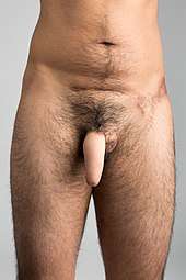
Phalloplasty
Phalloplasty is the construction or reconstruction of a penis, or the artificial modification of the penis by surgery. The term phalloplasty is also occasionally used to refer to penis enlargement.
| Phalloplasty | |
|---|---|
| Specialty | urology |
The first phalloplasty done for the purposes of sexual reassignment was performed on Michael Dillon, a trans man, in 1946 by Dr. Harold Gillies, which is documented in Pagan Kennedy's book The First Man-Made Man.
History
The Russian surgeon Nikolaj Bogoraz performed the first reconstruction of a total penis using rib cartilage in a reconstructed phallus made from a tubed abdominal flap in 1936.[1][2][3] The first female to male gender reassignment procedure was performed in 1946 by Sir Harold Gillies on fellow physician Michael Dillon, and his technique remained the standard one for decades. Later improvements in microsurgery made more techniques available.
Indications
A complete construction or reconstruction of a penis can be performed on patients who:
- Have congenital anomalies such as micropenis, epispadias, and hypospadias
- Have lost their penis
- Are trans men who desire sex reassignment surgery as part of their gender transition.
Techniques and related procedures
There are different techniques for phalloplasty. Construction of a new penis (sometimes called a neophallus) typically involves taking a tissue flap from a donor site (such as the forearm). Extending the urethra through the length of the neophallus is another goal of phalloplasty.[4]
Surgery for cisgender males is simpler than for female-to-male transgender patients, because the urethra requires less lengthening. The urethra of a trans man ends near the vaginal opening and has to be lengthened considerably. The lengthening of the urethra is where most complications occur.
With all types of phalloplasty in trans men, scrotoplasty can be performed using the labia majora (vulva) to form a scrotum where prosthetic testicles can be inserted. If vaginectomy, hysterectomy and/or oophorectomy have not been performed, they can be done at the same time.
Unlike metoidioplasty, phalloplasty requires an implanted erectile prosthesis to achieve an erection. This is usually done in a separate surgery to allow time for healing. There are several types of erectile prostheses, including malleable rod-like medical devices that allow the neo-penis to either stand up or hang down. Penile implants require a neophallus of appropriate length and volume in order to be a safe option. The long term success rates of implants in constructed penises are lower than the success rates of reconstruction in people born with penises. Good sensation in the reconstructed penis can help reduce the risk of the implant eventually eroding through the skin.[5]
Earlier techniques used a bone graft as part of reconstruction. Long-term follow-up studies from Germany and Turkey of more than 10 years proved that these reconstructions maintain their stiffness without later complications. Unfortunately, the reconstructed penis cannot become flaccid again without breaking the internal bone graft.
Temporary lengthening can also be gained by a procedure that releases the suspensory ligament where it is attached to the pubic bone, thereby allowing the penis to be advanced toward the outside of the body. The procedure is performed through a discreet horizontal incision located in the pubic region where the pubic hair will help conceal the incision site. However, scar formation can cause the penis to retract. Therefore, the American Urological Association "considers the division of the suspensory ligament of the penis for increasing penile length in adults to be a procedure which has not been shown to be safe or efficacious."[6]
As of November 2009, there is research in progress to synthesize corpora cavernosa (erectile tissue) in the lab on rabbits for eventual use in patients requiring penile construction surgery. Of the rabbits used in the preliminary studies, 8 of 12 had biological responses to sexual stimuli that was similar to the control, and four caused impregnation.[7]
Explanation of techniques
Flap from the arm
An operation using the forearm as a donor site is the easiest to perform but results in a cosmetically undesirable scar on the exposed area of the arm. Arm function may be hampered if the donor site does not heal properly. Electrolysis and/or laser hair reduction is required for a relatively hairless neophallus.
Sometimes a full-scale metoidioplasty is done a few months before the actual phalloplasty to reduce the possibility of complications after phalloplasty. Sensation is retained through the clitoral tissue at the base of the neophallus, and surgeons will often attempt to connect nerves together from the clitoris or nearby. Nerves from the flap and the tissue it has been attached to may eventually connect. This does not necessarily guarantee the ability to achieve genital orgasm after healing, as the most important task of nerve reconnection is to ensure the penis is able to sense injury, but it is rare to lose the ability to orgasm.
The following explanation of this technique has many similarities to other approaches, but the construction of the glans differs.
- The surgery starts (after the patient is prepped) with the forearm marked for graft size. After the graft is taken, another graft will be used to cover the arm (resulting in a secondary scar).
- The graft is dissected to expose the veins and antebrachial cutaneous nerves. (the latter done carefully for later reattachment)
- If the urethra is being constructed at the same time as the phallus, it is joined at this step. If not, the glans is shaped. Sometimes glansplasty is done in a separate surgical stage after urethral extension.
- A segment of vein going to the patient's groin is "borrowed" to allow easier joining of the graft with the preexisting tissues.
- The vein is carefully attached to the femoral artery.
- The blood supplies from the graft and the vein leading to the femoral artery are joined.
- The clitoral hood and ligament is cut away, and the nerve bundle is isolated for the time being. While this assumes the clitoral tissue is assimilated (buried) into the penis base, some surgeons give the option of leaving it as-is in a post metoidioplasty like state.
- The flap is partially attached physically while the surgeon attempts to join the nerve bundles.
- If the urethra was extended, it is now joined with a catheter that will remain in place for healing purposes for two to four weeks. Otherwise, the skin is sutured up and/or the scrotum is fabricated.
If the patient chooses to have the urethra extended to the glans of the neophallus, it is formed by the following steps:
- The labia minora is injected with a mixture of saline and epinephrine.
- It is then split open and layers separated using sharp and blunt dissection.
- The layers are wrapped around a catheter and stitched.
- A mucosal flap from the vagina may be used to bridge the urethra with the extension. This is often done in a separate procedure. Alternative graft locations include the mouth/cheeks or experimentally, the intestines. If the labia minora is not used during construction of the urethral extension, (or in the chance there is enough material remaining) it can be used during glansplasty to provide for better results compared with a full thickness skin graft.
Flap from the side of the chest
A relatively new technique involving a flap from the side of the chest under the armpit (known as a musculocutaneous latissimus dorsi free transfer flap) is a step forward in phalloplasty. The advantages of this technique over the older forearm flap technique include:
- Hairlessness (little to no electrolysis needed)
- Aesthetic appearance of normally colored skin (the glans may be tattooed to proper color)
- Capable of tactile sensation (as with any form of phalloplasty, this does not necessarily mean the ability to have a genital orgasm after healing, as the erogenous zone is limited to the base of the penis)
- Leaves an inconspicuous scar
- Has a lower occurrence of complications from both the initial surgery and the erectile prosthesis insertion
The disadvantageous include:
- Uses a motor nerve so erogenous sensation cannot be achieved, only tactile sensation.
- It can pull the nipple to the side causing it to be off the usual location.
This is a three-part surgery that takes place over a period of six to nine months. The steps consist of:
Neophallus creation using MLD free flap
- The surgery starts (after the patient is prepped) with the side of the chest marked for flap size.
- The flap is dissected to expose the veins and the thoracodorsal nerves.
- The flap, while still attached to the blood supply, is formed to a rough phallus shape by rolling the edges together.
- A segment of vein going to the patient's groin is "borrowed" to allow easier joining of the flap with the preexisting tissues.
- The vein is carefully attached to the femoral artery.
- The blood supplies from the flap and the vein leading to the femoral artery are joined.
- The clitoral hood and ligament are cut away and the nerve bundle is isolated.
- The flap is partially attached physically while the surgeon attempts to join the nerve bundles.
During initial recovery, the neophallus is protected from contact with other tissues with a specially constructed dressing as to avoid blood supply complications.
After three months, urethroplasty (urethral extension) is performed.
- The neophallus is dissected and a buccal (oral) mucosa graft inlaid into the created cavity and extended to the native urethra and joined to permanently allow urination while standing
- A catheter is placed for several weeks to allow for proper healing
After another three to six months, a device that allows an erection can be inserted.
Flap from the leg
The lower leg operation is similar to forearm flap with the exception that the donor scar is easily covered with a sock and/or pants and hidden from view. Other details are same as forearm flap, especially the need for permanent hair removal before the operation. A flap from the leg or another area where the scar is less noticeable may be combined with free forearm flap to create the urethral lengthening or to sculpt the glans penis.
Pubic area flap
The flap location is around the pelvic bone, usually running across the abdomen under the belly button. As such, there is a large horizontal scar that may not be aesthetically acceptable. The flap has a less natural appearance and may not maintain an erectile implant long term. Electrolysis is required before surgery with the alternative being clearing of hair via shaving, or chemical depilatory.
Gillies technique
This technique was pioneered by Sir Harold Delf Gillies as one of the first competent phalloplasty techniques. It was simply a flap of abdominal skin rolled into a tube to simulate a penis, with urethral extension being another section of skin to create a "tube within a tube." Early erectile implants consisted of a flexible rod. A later improvement involved the inclusion of a blood supply pedicle which was left in place to prevent tissue death before it was transplanted to the groin. Most latter techniques involve tissues with attached pedicle.
Abdominal muscle
Skin grafted muscle flaps have fallen from popularity. This procedure is a minimum of 3 steps and involves implantation of an expansion balloon to facilitate the amount of skin needed for grafting. The grafts have a less natural appearance and are less likely to maintain an erectile implant long term.
Subcutaneous soft silicone implant
This phalloplasty procedure involves the insertion of a subcutaneous soft silicone implant under the penile skin.[8][9][10][11]
No-touch surgical technique
This technique for penile prosthesis implantation is a surgical procedure developed by J. Francois Eid for the implantation of a penile prosthesis.[12] Implantation through the use of the "No-Touch" Technique minimizes the risk of infection.
As advancements in the design and manufacturing process of the IPP improved its mechanical survival infection has emerged as the leading cause of implant failure. Although relatively infrequent (varying from .06% to 8.9%) infection of a penile prosthesis results in serious medical consequences for the bearer, requiring complete removal of the device and permanent loss of penile size and anatomy.[13][14] Bacterial contamination of the device occurs during the surgery, and is caused by allowing direct or indirect contact of the prosthesis with the patient's skin. Over 70% of infections form from skin organisms including Staphylococcus epidermis, aureus, streptococcus and Candida albicans.[15]
Traditional strategies to combat infections aim at decreasing skin colony count such as scrubbing skin preparation with alcohol and chlorhexidine or kill bacteria once the implant is contaminated by skin flora such as intravenous antibiotics, antibiotic irrigation and antibiotic-coated implants. The "No-Touch" technique is unique in that it aims at preventing bacterial contamination of the prosthesis by completely eliminating contact of the device with the skin.[16]
Paired with the antibiotic-coated implant, the "No Touch" Technique decreases infection to a rate of 0.46%, opposing the traditional method which has an infection rate of 5%. The use of an antibiotic-coated implant and a no-touch surgical technique with skin preparation measures and peri-operative antibiotic use has been found to be of high importance in the prevention of infection among penile implants.[17] J. Francois Eid developed his no-touch technique in 2006 on the hypothesis that eliminating any contact between the prosthesis and the skin, either directly or indirectly via surgical instruments or gloves, should reduce the incidence of contamination of the device with skin flora responsible for infection.[15][18]
Procedure
Three days prior to the procedure, a patient is placed on oral fluoroquinolone, an antibacterial drug. During this time, the patient scrubs their lower abdomen and genitals daily with chlorhexidine soap. On the day of the surgery, Vancomycin and Gentamicin are administered intravenously one to two hours prior to the procedure. The lower abdomen and genitals are shaved, scrubbed for five minutes with a Chlorhexidine sponge and prepped with Chorhexidine/Alcohol applicator. The area is then draped with a surgical drape and a Vi Drape over the genitalia. Before the incision is made, a Foley catheter is inserted in the bladder through the urethra.
A 3-cm scrotal incision is made on the penoscrotal raphe and carried down through the subcutaneous tissue to the Buck's fascia. A Scott retractor, a flexible device that holds open the skin of the surgical site, is applied to the area.
Up until this stage of the surgery, the process has been consistent with the sanitary practices associated with standard surgical sterility.[19] At this stage of the "No-Touch" Technique, after the incision has been made, all instruments, including surgical gloves that have touched skin are discarded. A loose drape is then deployed over the entire surgical field and secured at the periphery with adhesive strips. A small opening in the drape is then made overlying the incision and yellow hooks utilized to secure the edges of the opening to the edges of the incision, completely covering and isolating the patient's skin. At this point new instruments and equipment are replaced and the entire prosthesis is inserted through the small opening of the loose drape. The loose drape allows for manipulation of the penis and scrotum required for this procedure without touching the skin.
Implantation of the device continues with an incision and dilation of corpora, sizing and placing the penile cylinders, and placement of the pump in the scrotum and the reservoir in the retropubic space. Saline is used throughout the implantation for irrigation. Once the corporotomies are closed and all of the tubing and components of the prosthesis covered with a layer of Buck's fascia, subcutaneous tissues are closed and the "No-Touch" drape is removed and skin closed.[12]
Future
In the future, bioengineering may be used to create fully functional penises.[20]
Common complications
As phalloplasty has improved over the decades, the risks and complications from surgery have been reduced. However, there is still a possibility of a need for revision surgery to repair incorrect healing.
A study of postoperative men showed that on average, 25% had one or more serious complications of the neopenis. The ones reported consisted of:
- Loss of the phallus from either disease or blood supply issues
- Cephalic vein thrombosis (blood clot)
- Arterial ischemia (shortage of blood supply)
- Infection
- Distal limited necrosis (death of parts of the penis)
- Hematoma (bruise)
In the same study, chances of complications of the extended urethra were higher, averaging 55%. The most common complications reported were:
- Urinary fistula (hole) requiring perineal urethrostomy
- Urinary fistula (hole) with conservative treatment
- Urinary retention (from stenosis or narrowing of the new urethra)
- (Erectile) prosthesis change (from complications)
- (Erectile) prosthesis explantation (removal of the prosthesis without replacement)
There is also a possibility of fat embolism "associated with liposuction and autologous fat transfer, a procedure where fat from liposuction is injected back into the same patient’s face, breast, buttocks or penis".[21]
See also
- List of transgender-related topics
- Penis enlargement
- Penis transplantation
- Sex reassignment surgery
References
- Rashid M, Tamimy MS (2013). "Phalloplasty: The dream and the reality". Indian J Plast Surg. 46 (2): 283–93. doi:10.4103/0970-0358.118606. PMC 3901910. PMID 24501465.
- Segal RL, Camper SB, Burnett AL (2014). "Modern utilization of penile prosthesis surgery: a national claim registry analysis". Int J Impot Res. 26 (5): 167–71. doi:10.1038/ijir.2014.11. PMID 24830674.
- Schultheiss D, Gabouev AI, Jonas U (2005). "Nikolaj A. Bogoraz (1874-1952): pioneer of phalloplasty and penile implant surgery". J Sex Med. 2 (1): 139–46. doi:10.1111/j.1743-6109.2005.20114.x. PMID 16422917.
- Morrison, Shane D.; Shakir, Afaaf; Vyas, Krishna S.; Kirby, Johanna; Crane, Curtis N.; Lee, Gordon K. (September 2016). "Phalloplasty: A Review of Techniques and Outcomes". Plastic and Reconstructive Surgery. 138 (3): 594–615. doi:10.1097/PRS.0000000000002518. ISSN 0032-1052. PMID 27556603.
- "Erectile Implants in Female-to-Male Transsexuals: Our Experience in 129 Patients" by Piet B. Hoebeke, Karel Decaestecker, Matthias Beysens, Yasmin Opdenakker, Nicolaas Lumen and Stan M. Monstrey, European Urology, February 2010
- "American Urological Association - Penile Augmentation Surgery". www.auanet.org. Retrieved 2018-03-14.
- "Bioengineered corporal tissue for structural and functional restoration of the penis" by Kuo-Liang Chen, Daniel Eberli, James J. Yoo, and Anthony Atala (Proceedings of the National Academy of Sciences, Vol. 106 No. 45, November 9, 2009)
- Shirvanian, V.; Lemperle, G.; Araujo Pinto, C.; Elist, J. J. (2014). "Shortened penis post penile prosthesis implantation treated with subcutaneous soft silicone penile implant: case report". International Journal of Impotence Research. 26 (3): 100–104. doi:10.1038/ijir.2013.44. PMID 24305609.
- "Shortened penis post penile prosthesis". MDLinx. Retrieved 16 February 2016.
- "A Retrospective Evaluation of the Safety and Effectiveness of a Silicone Block Implant for Elective Cosmetic Surgery of the Penis". SMSNA.org.
- Hannah Smothers (January 27, 2016). "Penis Implants Exist Now, and They Start at a Size Large". Cosmopolitan.com.
- Eid, J. Francois (2011). "No-Touch Technique". J Sex Med. 8 (1): 5–8. doi:10.1111/j.1743-6109.2010.02137.x. PMID 21199375.
- Mulcahy, John J. (2010). "Current approach to the treatment of penile implant infections". Therapeutic Advances in Urology. 2 (2): 69–75. doi:10.1177/1756287210370330. PMC 3126071. PMID 21789084.
- Gomelsky, A.; Dmochowski, RR (2003). "Antibiotic prophylaxis in urologic prosthetic surgery". Current Pharmaceutical Design. 9 (12): 989–96. doi:10.2174/1381612033455198. PMID 12678865.
- Eid, J. Francois; Wilson, Steven K.; Cleves, Mario; Salem, Emad A. (2012). "Coated Implants and "No Touch" Surgical Technique Decreases Risk of Infection in Inflatable Penile Prosthesis Implantation of 0.46%". Urology. 79 (6): 1310–5. doi:10.1016/j.urology.2011.11.076. PMID 22521187.
- Carson, CC (2004). "Efficacy of antibiotic impregnation of inflatable penile prostheses in decreasing infection in original implants". J Urol. 171 (4): 1611–4. doi:10.1097/01.ju.0000118245.66976.e1. PMID 15017233.
- Elmussareh, Muhammad; Goddard, Jonathan Charles; Summerton, Duncan John; Terry, Timothy Robin (2013). "Minimizing the risk of device infection in penile prosthetic surgery: a UK perspective". Journal of Clinical Urology. 6 (5): 280–288. doi:10.1177/2051415813488367.
- Muench, Peter J. (2013). "Infections Versus Penile Implants: The War on Bugs". Journal of Urology. 189 (5): 1631–1673. doi:10.1016/j.juro.2012.05.080. PMID 23085299.
- Richard Pearcy & Raj Persad, publication date unknown, "Inflatable penile prosthesis," in BJU International Website Atlas of Surgery and Surgical Devices, see , accessed 31 May 2014
- Chen KL, Eberli D, Yoo JJ, Atala A (November 2009). "Regenerative Medicine Special Feature: Bioengineered corporal tissue for structural and functional restoration of the penis". Proceedings of the National Academy of Sciences of the United States of America. 107 (8): 3346–50. doi:10.1073/pnas.0909367106. PMC 2840474. PMID 19915140.
- Zilg, Brita; Råsten-Almqvist, Petra (2017). "Fatal Fat Embolism After Penis Enlargement by Autologous Fat Transfer: A Case Report and Review of the Literature". Journal of Forensic Sciences. 62 (5): 1383–1385. doi:10.1111/1556-4029.13403. PMID 28749069.
Sources
- Total Phalloplasty Using a Musculocutaneous Latissimus Dorsi Flap by Sava V. Perovic, Rados Djinovic (British Journal of Urology, Reconstructive Urology, Volume 100 Issue 4, Sep 2007)
- New Technique of Total Phalloplasty With Reinnervated Latissimus Dorsi Myocutaneous Free Flap in Female-to-Male Transsexuals by Vesely, Jiri; Hyza, Petr; Ranno, Raul; Cigna, Emanuele; Monni, Nicola; al etc. (Annals of Plastic Surgery, Volume 58 Issue 5, May 2007)
- Simultaneous Penis and Perineum Reconstruction Using a Combined Latissimus Dorsi-Scapular Free Flap with Intraoperative Penile Skin Expansion by Rohrich, Rod J.; Allen, Terry; Lester, Fred; Young, Jonathan P.; Katz, Scott L. (Journal of Plastic and Reconstructive Surgery, Volume 99 Issue 4, April 1997)
- Neophalloplasty in Female-to-Male Transsexuals with the Island Tensor Fasciae Latae Flap by Santanelli, Fabio M.D., Ph.D.; Scuderi, Nicolò (Journal of Plastic and Reconstructive Surgery, Volume 105 Issue 6, May 2000)
- A New Surgical Procedure for Phallic Reconstruction: Istanbul Flap by Mutaf, Mehmet (Journal of Plastic and Reconstructive Surgery, Volume 105 Issue 4, April 2000)
- One-Stage Reconstruction of the Penis with Composite Iliac Crest and Lateral Groin Skin Flap by Sun, Guang-ci M.D.; Huang, Jin-jing (Annals of Plastic Surgery, Volume 15 Issue 6, December 1985)
- A Novel Single-Flap Technique for Total Penile Reconstruction: The Pedicled Anterolateral Thigh Flap by Lee, Gordon K.; Lim, Angeline F.; Bird, Erin (Journal of Plastic and Reconstructive Surgery, Volume 124 Issue 1, July 2009)
- Penile Reconstruction by the Free Scapular Flap and Malleable Penis Prosthesis by Yang, Mingyong; Zhao, Muxin; Li, Senkai; Li, Yangqun (Journal of Plastic and Reconstructive Surgery, Volume 59 Issue 1, July 2007)
- Hage, J. Joris (1992). Peniplastica Totalis to Reassignment Surgery of the External Genitalia in Female-to-Male Transsexuals. ISBN 978-90-5383-115-1.
- Long-Term Follow-Up of Total Penile Reconstruction with Sensate Osteocutaneous Free Fibula Flap in 18 Biological Male Patients by Sengezer, Mustafa; Öztürk, Serdar; Deveci, Mustafa; Odabasi, Zeki (Journal of Plastic and Reconstructive Surgery, Volume 114 Issue 2, August 2004)
- Long-Term Fate of the Bony Component in Neophallus Construction with Free Osteofasciocutaneous Forearm or Fibula Flap in 18 Female-to-Male Transsexuals by Papadopulos, Nikolaos A.; Schaff, Juergen; Biemer, Edgar (Journal of Plastic and Reconstructive Surgery, Volume 109 Issue 3, March 2002)
- Use of forearm free-flap phalloplasty in bladder exstrophy adults by Marc-Olivier Timsit, Pierre Mouriquand, Alain Ruffion, Alain Bouillot, Diala Dembelé, Arnaud Mejean, Fanny Lalloue, Albert Leriche and Nicolas Morel-Journel (BJU International, Volume 103 Issue 10, Dec 2008)
- Phalloplasty for female transsexuals with sensate free forearm flap by Rong-Hwang Fang, Jin-Teh Lin, Shiuh Ma (Microsurgery, Volume 15 Issue 5, Oct 2005)
- Long-term outcome of forearm flee-flap phalloplasty in the treatment of transsexualism by Albert Leriche, Marc-Olivier Timsit, Nicolas Morel-Journel, André Bouillot, Diala Dembele and Alain Ruffion (BJU International, Volume 101 Issue 10, Jan 2008)
- Penile Reconstruction: Is the Radial Forearm Flap Really the Standard Technique? by Monstrey, Stan; Hoebeke, Piet; Selvaggi, Gennaro; al etc. (Journal of Plastic and Reconstructive Surgery, Volume 124 Issue 2, August 2009)
- Addressing the ideal requirements by free flap phalloplasty: Some reflections on refinements of technique by J. Joris Hage, Floris H. De Graaf (Microsurgery, Volume 14 Issue 9, Oct 2005)
- Total Phallic Construction in Female to Male Transsexuals by Giulio Garaffa, Nim A. Christopher, David J. Ralph (Current Urology, Vol. 3, No. 3, 2009)
- Gender Reassigment by Dan Greenwald and Wayne Stadelmann (eMedicine Journal, Volume 2 Number 7, July 6, 2001)
- Gender Identity Disorders: Diagnostic and Surgical Aspects by Michael Sohn, and Hartmut Bosinski, MD (Journal of Sexual Medicine, Volume 4 Issue 5, Aug 2007)
- Glans sculpting in phalloplasty — experiences in female-to-male transsexuals by Rong-Hwang Fang, Yi-Sheng Kaoa, Shiuh Ma, Jin-Teh Lin (Journal of Plastic, Reconstructive & Aesthetic Surgery, Volume 51, Issue 5, July 1998)
- Severe Penile Injuries: Etiology, Management and Outcomes by Sava V. Perovic, Urologia Polska (Polish Journal of Urology) 2005/58/3, ISSN 0500-7208.