Peroneus longus
In human anatomy, the peroneus longus (also known as fibularis longus) is a superficial muscle in the lateral compartment of the leg, and acts to evert and plantarflex the ankle.
| Fibularis longus | |
|---|---|
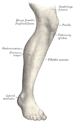 Lateral aspect of right leg | |
 Peroneus longus labeled at right | |
| Details | |
| Origin | Proximal part of lateral surface of shaft of fibula[1] |
| Insertion | First metatarsal, medial cuneiform[1] |
| Artery | fibular (peroneal) artery |
| Nerve | Superficial fibular (peroneal) nerve[1] |
| Actions | plantarflexion, eversion, support arches[1] |
| Antagonist | Tibialis anterior muscle, T.A. does Inversion and Dorsiflexion |
| Identifiers | |
| Latin | Musculus peronaeus longus, musculus fibularis longus |
| TA | A04.7.02.041 |
| FMA | 22539 |
| Anatomical terms of muscle | |
The muscle, the longest and most superficial of the three peroneus muscles, is attached proximally to the head of the fibula and its 'belly' runs down most of this bone. It becomes a tendon that goes posteriorly around the lateral malleolus of the ankle, then continues under the foot to attach to the medial cuneiform and first metatarsal. It is innervated by the Superficial peroneal nerve (L5,S1)
Structure
It arises from the head and upper two-thirds of the lateral surface of the body of the fibula, from the deep surface of the fascia, and from the intermuscular septa between it and the muscles on the front and back of the leg; occasionally also by a few fibers from the lateral condyle of the tibia. Between its attachments to the head and to the body of the fibula there is a gap through which the common peroneal nerve passes to the front of the leg.[2]
It ends in a long tendon, which runs behind the lateral malleolus, in a groove common to it and the tendon of the peroneus brevis; the groove is converted into a canal by the superior peroneal retinaculum, and the tendons in it are contained in a common mucous sheath.[2]
The tendon then extends obliquely forward across the lateral side of the foot, below the peroneal tubercle, and the tendon of the peroneus brevis, and under cover of the inferior peroneal retinaculum.[2]
It crosses the lateral side of the cuboid, and then runs on the under surface of that bone in a groove which is converted into the peroneal canal by the long plantar ligament; the tendon then crosses the sole of the foot obliquely, and is inserted into the lateral side of the base of the first metatarsal bone and the lateral side of the medial cuneiform.[2]
Occasionally it sends a slip to the base of the second metatarsal bone.[2]
The tendon changes its direction at two points: first, behind the lateral malleolus; secondly, on the cuboid bone; in both of these situations the tendon is thickened, and, in the latter, a sesamoid fibrocartilage (sometimes a bone), is usually developed in its substance.[2]
Function
The peroneus longus and brevis muscles plantarflex the foot, in conjunction with the tibialis posterior, antagonizing the tibialis anterior and peroneus tertius, which are dorsiflexors of the foot.[2]
The peroneus longus also everts the sole of the foot, and from the oblique direction of the tendon across the sole of the foot is an important agent in the maintenance of the transverse arch.[2]
Taking their fixed points below, the peroneus muscles serve to steady the leg upon the foot.[2]
This is especially the case in standing upon one leg, when the tendency of the superincumbent weight is to throw the leg medialward; the peroneus longus overcomes this tendency by drawing on the lateral side of the leg.[2]
History
Etymology
The terms Peroneus (i.e., Longus and Brevis) and Peroneal (i.e., Artery, Retinaculum) are derived from the Greek word Perone (pronounced Pair-uh-knee) meaning pin of a brooch or a buckle. In medical terminology, both terms refer to being of or relating to the fibula or to the outer portion of the leg.
Additional images
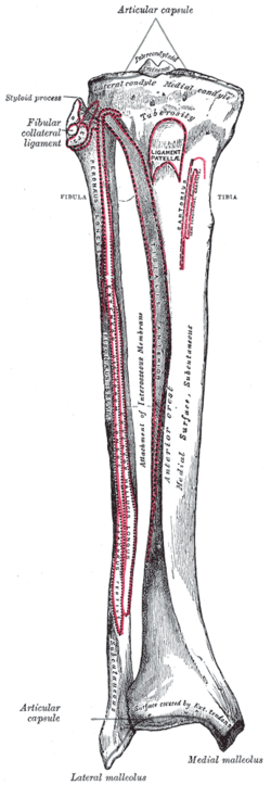 Bones of the right leg. Anterior surface
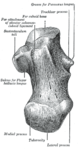 Left calcaneus, inferior surface
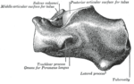 Left calcaneus, lateral surface
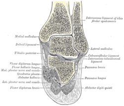 Coronal section through right talocrural and talocalcaneal joints
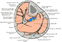 Cross-section through middle of leg
 The popliteal, posterior tibial, and fibular arteries
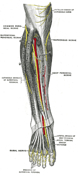 Deep nerves of the front of the leg
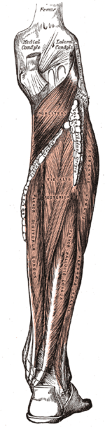
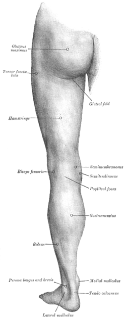 Back of left lower extremity
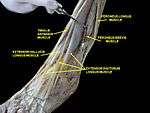 Musclea of Leg (lateral view, deep dissection)
See also
References
This article incorporates text in the public domain from page 486 of the 20th edition of Gray's Anatomy (1918)
- "Peroneus longus". Loyola University Chicago. Retrieved 4 August 2019.
- Gray's Anatomy (1918), see infobox
| Authority control |
|---|