Peripheral venous catheter
In medicine, a peripheral venous catheter (PVC), peripheral venous line or peripheral venous access catheter is a catheter (small, flexible tube) placed into a peripheral vein for venous access to administer intravenous therapy such as medication fluids.

1. The catheter itself is composed of (a) a tip for insertion into the vein, (b) wings for manual handling and securing the catheter with adhesives, (c) a valve to allow injection of drugs with a syringe, (d) an end which allows connection to an intravenous infusion line, and capping in between uses.
2. The needle (partially retracted) which serves only as a guidewire for inserting the cannula.
3. The protection cap which is removed before use.
Use
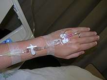
The catheter is introduced into the vein by a needle (similar to blood drawing), which is subsequently removed while the small plastic cannula remains in place. The catheter is then fixed by taping it to the patient's skin or using an adhesive dressing.
A peripheral venous catheter is the most commonly used vascular access in medicine. It is given to most emergency department and surgical patients, and before some radiological imaging techniques using radiocontrast, for example. In the United States, in the 1990s, more than 25 million patients had a peripheral venous line each year.[1]
A peripheral venous catheter is usually placed in a vein on the hand or arm. It should be distinguished from a central venous catheter which is inserted in a central vein (usually in the internal jugular vein of the neck or the subclavian vein of the chest), or an arterial catheter which can be placed in a peripheral or central artery. In children, a topical anaesthetic gel (such as lidocaine) may be applied to the insertion site to facilitate placement.
Complications
Infection, phlebitis, extravasation, infiltration, air embolism, hemorrhage (bleeding) and formation of a hematoma (bruise) may occur. Because of the risk of insertion-site infection the CDC advises in their guideline that the catheter needs to be replaced every 96 hours.[2] However, the need to replace these catheters routinely is debated.[3] Expert management has been shown to reduce the complications of peripheral lines.[1][4]
Sizes
Sizes of peripheral venous catheters can be given by Birmingham gauge or French gauge. Diameter is proportional to French gauge and inversely proportional to Birmingham gauge.
| Birmingham gauge | Diameter (mm) | Maximum flow rate (ml/min)[5] | Color[5] |
|---|---|---|---|
| 26 | 0.46 | 13-15 | Black |
| 24 | 0.60 | 36 | Yellow |
| 22 | 0.90 | 56 | Blue |
| 20 | 1.10 | 40-80 | Pink |
| 18 | 1.30 | 75-120 | Green |
| 17 | 1.50 | 128-133 | White |
| 16 | 1.80 | 236 | Grey |
| 14 | 2.00 | 270 | Orange |
History
The insertion of a plastic cannula and withdrawal of the needle was introduced as a technique in 1945.[6] The first disposable version to be marketed was the Angiocath, first sold in 1964. In the 1970s and 1980s, the use of plastic cannulas became routine, and the insertion changed from being the responsibility of the doctor, to being the responsibility of a nurse.[7]
Newer catheters have been equipped with additional safety features to avoid needlestick injuries. Modern catheters consist of synthetic polymers such as teflon (hence the often used term 'Venflon' or 'Cathlon' for these venous catheters). In 1950 they consisted of PVC plastic.[8][9] In 1983, the first polyurethane version was introduced.[7]
Additional images
 An arm board is recommended for immobilizing the extremity for cannulation of the hand, the foot or the antecubital fossa in children.[10]
 Just before inserting.
 The catheter in between uses.
 Newer catheter with additional safety features.
References
- Soifer NE, Borzak S, Edlin BR, Weinstein RA (March 1998). "Prevention of peripheral venous catheter complications with an intravenous therapy team: a randomized controlled trial". Arch. Intern. Med. 158 (5): 473–77. doi:10.1001/archinte.158.5.473. PMID 9508225.
- CDC Morbidity and Mortality Weekly Report Aug 2002. "Guidelines for the Prevention of Intravascular Catheter-Related Infections". Retrieved 2008-03-13.
- Bregenzer T, Conen D, Sakmann P, Widmer AF (January 1998). "Is routine replacement of peripheral intravenous catheters necessary?". Arch. Intern. Med. 158 (2): 151–56. doi:10.1001/archinte.158.2.151. PMID 9448553.
- Miller JM, Goetz AM, Squier C, Muder RR (1996). "Reduction in nosocomial intravenous device-related bacteremias after institution of an intravenous therapy team". J Intraven Nurs. 19 (2): 103–06. PMID 8852171.
- p. 110 in: Edward Doyle (2007). Paediatric Anaesthesia. OUP Oxford. ISBN 9780199202799.
- Narr, Bradly J.; Southorn, Peter A. (1 October 2008). "The Massa or Rochester Plastic Needle". Mayo Clinic Proceedings. 83 (10): 1165–1167. doi:10.4065/83.10.1165. ISSN 0025-6196. Retrieved 16 April 2019.
- Rivera AM, Strauss KW, Van Zundert A, Mortier E (2005). "The history of peripheral intravenous catheters : How little plastic tubes revolutionized medicine". Acta Anaesthesiol. Belg.
- Massa DJ; Lundy JS; Faulconer A, Jr; Ridley RW (5 Jul 1950). "A plastic needle". Proc Staff Meet Mayo Clin. 25 (14): 413–15. PMID 15430460.
- Strauss KW, Onia R, Van Zundert A (August 2008). "Peripheral intravenous catheter use in Europe: Towards the use of safety devices". Acta Anaesthesiologica Scandinavica. 52 (6): 798–804. doi:10.1111/j.1399-6576.2008.01664.x. hdl:1854/LU-529472. PMID 18477072.
- p. 349 in: James R. Roberts, Jerris R. Hedges (2013). Roberts and Hedges' Clinical Procedures in Emergency Medicine E-Book (6 ed.). Elsevier Health Sciences. ISBN 9781455748594.