Periapical periodontitis
Periapical periodontitis (AP) is an acute or chronic inflammatory lesion around the apex of a tooth root which is usually caused by bacterial invasion of the pulp of the tooth.[1] The term is derived from peri- meaning "around", apical referring to the apex of the root (the tip of the root), and -itis meaning a disease characterized by inflammation. Periapical periodontitis can be considered a sequela in the natural history of dental caries (tooth decay), irreversible pulpitis and pulpal necrosis, since it is the likely outcome of untreated dental caries, although not always. In some cases, periapical periodontitis can occur due to occlusal high spots post-restoration, endodontic root filling material extrusion or bacterial invasion and infection from a gingival communication (rather than a pulpal source). Periapical periodontitis may develop into a periapical abscess, where a collection of pus forms at the end of the root, the consequence of spread of infection from the tooth pulp (odontogenic infection), or into a periapical cyst, where an epithelial lined, fluid filled structure forms.
| Periapical periodontitis | |
|---|---|
| Other names | Apical periodontitis, periradicular periodontitis |
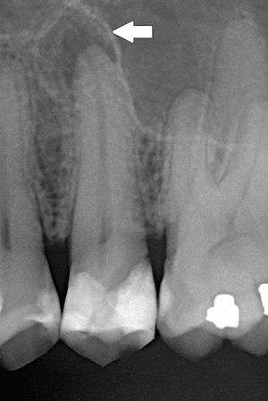 | |
| Periapical dental radiograph showing chronic periapical periodontitis on the root of the left maxillary second premolar. Note large restoration present in the tooth, which will have undergone pulpal necrosis at some point before the development of this lesion. | |
| Specialty | Dentistry |
Diagnosis
The radiographic features of periapical inflammatory lesions vary depending on the time course of the lesion. Because very early lesions may not show any radiographic changes, diagnosis of these lesions relies solely on the clinical symptoms. More chronic lesions may show lytic (radiolucent) or sclerotic (radiopaque) changes, or both.
Classification
The type of periapical periodontitis is usually classified according to whether it is an acute/symptomatic process or a chronic/asymptomatic process.
Acute periapical periodontitis
Acute periapical periodontitis, also termed acute apical periodontitis, acute periradicular periodontitis, or symptomatic periapical periodontitis.
Chronic periapical periodontitis
Chronic periapical periodontitis, also termed chronic apical periodontitis, chronic periradicular periodontitis, or asymptomatic periapical periodontitis
A periapical granuloma (also termed an apical granuloma or a radicular granuloma) is mass of chronically inflamed granulation tissue that forms at the apex of the root of a nonvital (dead) tooth.[2] However, a periapical granuloma does not contain granulomatous inflammation, and therefore is not a true granuloma, but the term periapical granuloma is in common use.[2]
Treatment
Treatment options include antibiotic therapy (not a permanent solution), endodontic (root canal) therapy, or extraction.
Epidemiology
Periapical periodontitis of some form is a very common condition. The prevalence of periapical periodontitis is generally reported to vary according to age group, e.g. 33% in those aged 20–30, 40% in 30- to 40-year-olds, 48% in 40- to 50-year-olds, 57% in 50- to 60-year-olds and 62% in those over the age of 60.[3] Most epidemiologic data has been generated in European countries, especially Scandinavia. Millions of root canal treatments are carried out in the United States each year, although the total number of root canal treatments is an imperfect indicator of the prevalence of periapical periodontitis, since not always is it performed due to the presence of periapacial periodontitis, and not all cases of asymptomatic periodontitis will be treated in this manner, either due to lack of patient attendance or watchful waiting.
References
- Segura-Egea, JJ.; Castellanos-Cosano, L.; Machuca, G.; Lopez-Lopez, J.; Martin-Gonzalez, J.; Velasco-Ortega, E.; Sanchez-Dominguez, B.; Lopez-Frias, FJ. (1 January 2012). "Diabetes mellitus, periapical inflammation and endodontic treatment outcome". Medicina Oral Patología Oral y Cirugia Bucal. 17 (2): e356–e361. doi:10.4317/medoral.17452. PMC 3448330. PMID 22143698.
- Neville BW, Damm DD, Allen CA, Bouquot JE (2002). Oral & maxillofacial pathology (2nd ed.). Philadelphia: W.B. Saunders. pp. 113–124. ISBN 978-0721690032.
- Hargreaves KM; Cohen S, eds. (2010). Cohen's pathways of the pulp. Berman LH (web editor) (10th ed.). St. Louis, Mo.: Mosby Elsevier. pp. 529–555. ISBN 978-0-323-06489-7.
External links
| Classification |
|
|---|