Percutaneous nephrostomy
Percutaneous nephrostomy is an interventional radiology/surgical procedure in which the renal pelvis is punctured whilst using imaging as guidance. Images are obtained once an antegrade pyelogram (an injection of contrast), with a fine needle, has been performed. This contrast is used to show calcifications at the renal pelvis. A nephrostomy tube may then be placed to allow drainage.[2]
| Percutaneous nephrostomy | |
|---|---|
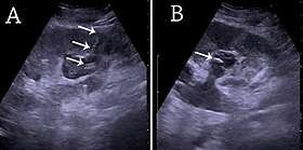 (A) Renal ultrasonograph of percutaneous nephrostomy tube placed through a calyx in the lower pole of a kidney with hydronephrosis. (B) The pigtail catheter is placed in the dilated calyx. The tube in (A) and the pigtail in (B) are marked with white arrows.[1] | |
| ICD-9-CM | 55.03 |
| OPS-301 code | 5-550 |
See also
References
- Content initially copied from: Hansen, Kristoffer; Nielsen, Michael; Ewertsen, Caroline (2015). "Ultrasonography of the Kidney: A Pictorial Review". Diagnostics. 6 (1): 2. doi:10.3390/diagnostics6010002. ISSN 2075-4418. PMC 4808817. (CC-BY 4.0)
- Longmore, Murray; Ian Wilkinson; Tom Turmezei; Chee Kay Cheung (2007). Oxford Handbook of Clinical Medicine, 7th edition. Oxford University Press. p. 731. ISBN 0-19-856837-1.
This article is issued from
Wikipedia.
The text is licensed under Creative
Commons - Attribution - Sharealike.
Additional terms may apply for the media files.