Parafollicular cell
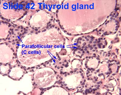
Parafollicular cells, also called C cells, are neuroendocrine cells in the thyroid. The primary function of these cells is to secrete calcitonin. They are located adjacent to the thyroid follicles and reside in the connective tissue. These cells are large and have a pale stain compared with the follicular cells. In teleost and avian species these cells occupy a structure outside the thyroid gland named the ultimobranchial body.
| Parafollicular cell | |
|---|---|
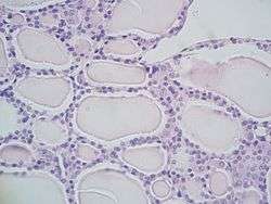 Microscopic section of the thyroid showing follicles, where parafollicular cells reside | |
| Details | |
| Location | Thyroid |
| Function | Calcitonin secretion |
| Identifiers | |
| TH | H3.08.02.4.00009 |
| Anatomical terms of microanatomy | |
Structure
Parafollicular cells are pale-staining cells found in small number in the thyroid and are typically situated basally in the epithelium, without direct contact with the follicular lumen. They are always situated within the basement membrane, which surrounds the entire follicle.
Development
Parafollicular cells are derived from pharyngeal endoderm.[1][2] Embryologically, they associate with the ultimobranchial body, which is a ventral derivative of the fourth (or fifth) pharyngeal pouch. Parafollicular cells were previously believed to be derived from the neural crest based on a series of experiments in quail-chick chimeras.[3][4] However, lineage tracing experiments in mice revealed that parafollicular cells are derived from the endoderm origin.[5]
Function
Parafollicular cells secrete calcitonin, a hormone that participates in the regulation of calcium metabolism. It is important in fish and rodents, but its relevance in human calcium homeostasis has not been demonstrated. Calcitonin lowers blood levels of calcium by inhibiting the resorption of bone by osteoclasts, and its secretion is increased proportionally with the concentration of calcium.[6]
Parafollicular cells are also known to secrete in smaller quantities several neuroendocrine peptides such as serotonin, somatostatin or CGRP.[7][8][9] They may also have a role in regulating thyroid hormones production locally, as they express thyrotropin-releasing hormone.[10][11]
Clinical significance
When parafollicular cells become cancerous, they lead to medullary carcinoma of the thyroid.
References
- Nilsson M, Williams D (July 2016). "On the Origin of Cells and Derivation of Thyroid Cancer: C Cell Story Revisited". European Thyroid Journal. 5 (2): 79–93. doi:10.1159/000447333. PMC 4949372. PMID 27493881.
- Johansson, E., Andersson, L., Örnros, J., Carlsson, T., Ingeson-Carlsson, C., Liang, S., … Nilsson, M. (2015). Revising the embryonic origin of thyroid C cells in mice and humans. Development, 142(20), 3519–3528. http://doi.org/10.1242/dev.126581
- Le Douarin N, Fontaine J, Le Lièvre C (March 1974). "New studies on the neural crest origin of the avian ultimobranchial glandular cells--interspecific combinations and cytochemical characterization of C cells based on the uptake of biogenic amine precursors". Histochemistry. 38 (4): 297–305. doi:10.1007/bf00496718. PMID 4135055.
- Barasch J, Gershon MD, Nunez EA, Tamir H, al-Awqati Q (December 1988). "Thyrotropin induces the acidification of the secretory granules of parafollicular cells by increasing the chloride conductance of the granular membrane". The Journal of Cell Biology. 107 (6 Pt 1): 2137–47. doi:10.1083/jcb.107.6.2137. PMC 2115661. PMID 2461947.
- Johansson E, Andersson L, Örnros J, Carlsson T, Ingeson-Carlsson C, Liang S, Dahlberg J, Jansson S, Parrillo L, Zoppoli P, Barila GO, Altschuler DL, Padula D, Lickert H, Fagman H, Nilsson M (October 2015). "Revising the embryonic origin of thyroid C cells in mice and humans". Development. 142 (20): 3519–28. doi:10.1242/dev.126581. PMC 4631767. PMID 26395490.
- Melmed S, Polonsky KS, Larsen PR, Kronenberg HM (2011). Williams Textbook of Endocrinology (12th ed.). Saunders. pp. 1250–1252. ISBN 978-1437703245.
- Zabel M (December 1984). "Ultrastructural localization of calcitonin, somatostatin and serotonin in parafollicular cells of rat thyroid". The Histochemical Journal. 16 (12): 1265–72. doi:10.1007/bf01003725. PMID 6152264.
- Barasch JM, Mackey H, Tamir H, Nunez EA, Gershon MD (September 1987). "Induction of a neural phenotype in a serotonergic endocrine cell derived from the neural crest" (PDF). The Journal of Neuroscience. 7 (9): 2874–83. doi:10.1523/JNEUROSCI.07-09-02874.1987. PMID 3305802.
- Bernd P, Gershon MD, Nunez EA, Tamir H (March 1981). "Separation of dissociated thyroid follicular and parafollicular cells: association of serotonin binding protein with parafollicular cells". The Journal of Cell Biology. 88 (3): 499–508. doi:10.1083/jcb.88.3.499. PMC 2112761. PMID 7217200.
- Gkonos PJ, Tavianini MA, Liu CC, Roos BA (December 1989). "Thyrotropin-releasing hormone gene expression in normal thyroid parafollicular cells". Molecular Endocrinology. 3 (12): 2101–9. doi:10.1210/mend-3-12-2101. PMID 2516877.
- Morillo-Bernal J, Fernández-Santos JM, Utrilla JC, de Miguel M, García-Marín R, Martín-Lacave I (August 2009). "Functional expression of the thyrotropin receptor in C cells: new insights into their involvement in the hypothalamic-pituitary-thyroid axis". Journal of Anatomy. 215 (2): 150–8. doi:10.1111/j.1469-7580.2009.01095.x. PMC 2740962. PMID 19493188.
Further reading
- Kameda Y (October 1987). "Localization of immunoreactive calcitonin gene-related peptide in thyroid C cells from various mammalian species". The Anatomical Record. 219 (2): 204–12. doi:10.1002/ar.1092190214. PMID 3120623.
- Kameda Y, Nishimaki T, Miura M, Jiang SX, Guillemot F (January 2007). "Mash1 regulates the development of C cells in mouse thyroid glands". Developmental Dynamics. 236 (1): 262–70. doi:10.1002/dvdy.21018. PMID 17103415.
- Kameda Y, Nishimaki T, Chisaka O, Iseki S, Sucov HM (October 2007). "Expression of the epithelial marker E-cadherin by thyroid C cells and their precursors during murine development". The Journal of Histochemistry and Cytochemistry. 55 (10): 1075–88. doi:10.1369/jhc.7a7179.2007. PMID 17595340.
- Kameda Y, Ito M, Nishimaki T, Gotoh N (March 2009). "FRS2alpha is required for the separation, migration, and survival of pharyngeal-endoderm derived organs including thyroid, ultimobranchial body, parathyroid, and thymus". Developmental Dynamics. 238 (3): 503–13. doi:10.1002/dvdy.21867. PMID 19235715.
- Kameda Y (March 2016). "Cellular and molecular events on the development of mammalian thyroid C cells". Developmental Dynamics. 245 (3): 323–41. doi:10.1002/dvdy.24377. PMID 26661795.
- Baber EC (1876). "Contributions to the Minute Anatomy of the Thyroid Gland of the Dog". Philosophical Transactions of the Royal Society of London. 166: 557–568. doi:10.1098/rstl.1876.0021. JSTOR 109205.
External links
- Histology image: 42_04 at the University of Oklahoma Health Sciences Center
- Histology image: 14302loa – Histology Learning System at Boston University
- Histology at KUMC endo-/endo10
- Anatomy Atlases - Microscopic Anatomy, plate 15.287
| Authority control |
|---|