Paneth cell
Paneth cells are a principal cell type of the small intestine epithelium, along with goblet cells, enterocytes, and enteroendocrine cells.[1] A few may also be found sporadically in the cecum and appendix. They are identified microscopically by their location just below the intestinal stem cells in the intestinal glands (also called crypts of Lieberkühn) and the large eosinophilic refractile granules that occupy most of their cytoplasm. Paneth cells are named after Joseph Paneth (1857–1890), an Austrian physiologist.
| Paneth cell | |
|---|---|
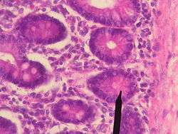 Paneth cells | |
| Details | |
| Location | Small intestine epithelium |
| Identifiers | |
| Latin | cellula panethensis |
| MeSH | D019879 |
| TH | H3.04.03.0.00017 |
| Anatomical terms of microanatomy | |
These granules consist of several anti-microbial compounds and other compounds that are known to be important in immunity and host-defense. When exposed to bacteria or bacterial antigens, Paneth cells secrete some of these compounds into the lumen of the intestinal gland, thereby contributing to maintenance of the gastrointestinal barrier.
Structure
Paneth cells are found throughout the small intestine and the appendix at the base of the intestinal glands.[2] The Paneth cell numbers demonstrate an ascending trend with highest numbers towards the distal end of the small intestine. Like the other epithelial cell lineages in the small intestine, Paneth cells originate at the stem cell region near the bottom of the gland.[3] However, unlike the other epithelial cell types, Paneth cells migrate downward from the stem cell region and settle just adjacent to it.[3] This close relationship to the stem cell region is thought to suggest that Paneth cells are important in defending the gland stem cells from microbial damage,[3] although their function is not entirely known.[2] Furthermore, among the four aforementioned intestinal cell lineages, the Paneth cells live the longest (18–23 days).
Function
"These cells synthesize and secrete substantial quantities of antimicrobial peptides and proteins. More recent studies have determined that these antimicrobial molecules are key mediators of host-microbe interactions, including homeostatic balance with colonizing microbiota and innate immune protection from enteric pathogens. Perhaps more intriguing, Paneth cells secrete factors that help sustain and modulate the epithelial stem and progenitor cells that cohabitate in the crypts and rejuvenate the small intestinal epithelium."[4]
Small intestinal crypts house stem cells that serve to constantly replenish epithelial cells that die and are lost from the villi.
Protection of these stem cells is essential for long-term maintenance of the intestinal epithelium, and the location of Paneth cells adjacent to stem cells suggests that they play a critical role in defending epithelial cell renewal.
Sensing microbiota
Paneth cells sense bacteria via MyD88-dependent toll-like receptor (TLR) activation which then triggers antimicrobial action.[5]
Secretions
The principal defense molecules secreted by Paneth cells are alpha-defensins, which are known as cryptdins in mice.[6] These peptides have hydrophobic and positively charged domains that can interact with phospholipids in cell membranes. This structure allows defensins to insert into membranes, where they interact with one another to form pores that disrupt membrane function, leading to cell lysis. Due to the higher concentration of negatively charged phospholipids in bacterial than vertebrate cell membranes, defensins preferentially bind to and disrupt bacterial cells, sparing the cells they are functioning to protect.[7]
Paneth cells are stimulated to secrete defensins when exposed to bacteria (both Gram positive and negative types) or such bacterial products as lipopolysaccharide, muramyl dipeptide and lipid A.
In addition to defensins, Paneth cells secrete lysozyme,[8] tumor necrosis factor-alpha,[8] and phospholipase A2. Lysozyme and phospholipase A2 both have clear antimicrobial activity. This battery of secretory molecules gives Paneth cells a potent arsenal against a broad spectrum of agents, including bacteria, fungi and even some enveloped viruses.
References
- Horst Ibelgaufts. "Go to Cells-Talk.com". Copewithcytokines.org. Retrieved 2016-09-17.
- "Paneth's cell | anatomy". Britannica.com. Retrieved 2016-09-17.
- Christopher Duggan; John B. Watkins; W. Allan Walker. Nutrition in Pediatrics: Basic Science, Clinical Applications. Books.google.com. p. 244. Retrieved 2016-09-17.
- Clevers, HC (2013). "Paneth cells: maestros of the small intestinal crypts". Cite journal requires
|journal=(help) - Vaishnava, S; Behrendt, CL; Ismail, AS; Eckmann, L; Hooper, LV (Dec 30, 2008). "Paneth cells directly sense gut commensals and maintain homeostasis at the intestinal host-microbial interface". Proceedings of the National Academy of Sciences of the United States of America. 105 (52): 20858–63. doi:10.1073/pnas.0808723105. PMC 2603261. PMID 19075245.
- Wilson C, Ouellette A, Satchell D, Ayabe T, López-Boado Y, Stratman J, Hultgren S, Matrisian L, Parks W (1999). "Regulation of intestinal alpha-defensin activation by the metalloproteinase matrilysin in innate host defense". Science. 286 (5437): 113–7. doi:10.1126/science.286.5437.113. PMID 10506557.
- Ayabe T, Satchell D, Wilson C, Parks W, Selsted M, Ouellette A (2000). "Secretion of microbicidal alpha-defensins by intestinal Paneth cells in response to bacteria". Nat Immunol. 1 (2): 113–8. doi:10.1038/77783. PMID 11248802.
- Kierszenbaum, Abraham L. (2002). Histology and cell biology : an introduction to pathology. St. Louis [u.a.]: Mosby. p. 434. ISBN 0-323-01639-1.
- Bibliography
- Ganz T (1999). "Defensins and host defense". Science. 286 (5439): 420–1. doi:10.1126/science.286.5439.420. PMID 10577203.
- Ganz T (2000). "Paneth cells--guardians of the gut cell hatchery". Nat Immunol. 1 (2): 99–100. doi:10.1038/77884. PMID 11248797.
External links
- Histology image: 11604loa – Histology Learning System at Boston University - "Endocrine System: duodenum, enteroendocrine cells"
- Histology image: 11606loa – Histology Learning System at Boston University - "Digestive System: Alimentary Canal - duodenum, Paneth cells"
- Overview and diagram at colostate.edu
- Histology at ucsd.edu
| Authority control |
|---|