Oxyphil cell (parathyroid)
In the parathyroid gland, the parathyroid oxyphil cell is larger and lighter staining than the parathyroid chief cell.[1]
| Oxyphil cell | |
|---|---|
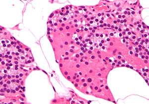 High magnification micrograph of parathyroid. H&E stain. The cells with orange/pink staining cytoplasm are oxyphil cells | |
| Details | |
| Location | Parathyroid gland |
| Identifiers | |
| TH | H3.08.02.5.00005 |
| Anatomical terms of microanatomy | |
These cells can be found in clusters in the center of the section and at the periphery.[2][3][4][5] Oxyphil cells appear at the onset of puberty, but have no known function. With nuclear medicine scans, they selectively take up the Technetium-sestamibi complex radiotracer to allow delineation of glandular anatomy.[6] Oxyphil cells have been shown to express parathyroid-relevant genes found in the chief cells and have the potential to produce additional autocrine/paracrine factors, such as parathyroid hormone-related protein (PTHrP) and calcitriol.[7] More work needs to be done to fully understand the functions of these cells and their secretions.
References
- Histology image:15002loa from Vaughan, Deborah (2002). A Learning System in Histology: CD-ROM and Guide. Oxford University Press. ISBN 978-0195151732.
- Gartner, p. 208, Fig. 3
- Ross, p. 628, Fig. 1
- DiFiore, pp. 270 - 271
- Wheater, pp. 312 - 313
- "Minimally Invasive Radio-guided Surgery for Primary Hyperparathyroidism," Annals of Surgical Oncology 12/07 14(12) pp 3401-3402
- Ritter, Haughey, Miller, Brown (2012). "Differential gene expression by oxyphil and chief cells of human parathyroid glands". J Clin Endocrinol Metab. 97 (8): E1499–505. doi:10.1210/jc.2011-3366. PMC 3591682. PMID 22585091.CS1 maint: multiple names: authors list (link)
| Authority control |
|---|