Osteosclerosis
Osteosclerosis is a disorder that is characterized by abnormal hardening of bone and an elevation in bone density. It may predominantly affect the medullary portion and/or cortex of bone. Plain radiographs are a valuable tool for detecting and classifying osteosclerotic disorders.[1][2] It can manifest in localized or generalized osteosclerosis. Localized osteosclerosis can be caused by Legg–Calvé–Perthes disease, sickle-cell disease and osteoarthritis among others. Osteosclerosis can be classified in accordance with the causative factor into acquired and hereditary.[2][1]
| Osteosclerosis | |
|---|---|
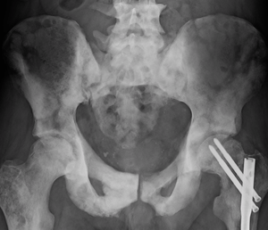 | |
| Sclerosis of the bones of the pelvis due to prostate cancer metastases | |
| Specialty | Medical genetics |
Types
Acquired osteosclerosis
- Osteogenic bone metastasis caused by carcinoma of prostate and breast
- Paget's disease of bone
- Myelofibrosis (primary disorder or secondary to intoxication or malignancy)
- Osteosclerosing types of chronic osteomyelitis
- Hypervitaminosis D
- hypoparathyroidism
- Schnitzler syndrome[3]
- Monoclonal IgM Kappa cryoglobulinemia[4]
- Hepatitis C.[5]
Hereditary osteosclerosis
- Malignant infantile osteopetrosis[6]
- Neuropathic infantile osteopetrosis
- Infantile osteopetrosis with renal tubular acidosis
- Infantile osteopetrosis with immunodeficiency
- IO with leukocyte adhesion deficiency syndrome (LAD-III)
- Intermediate osteopetrosis
- Autosomal dominant osteopetrosis (Albers-Schonberg)
- Pyknodysostosis (osteopetrosis acro-osteolytica)[2]
- Osteopoikilosis (Buschke–Ollendorff syndrome)[2]
- Osteopathia striata with cranial sclerosis[2]
- Mixed sclerosing bone dysplasia
- Progressive diaphyseal dysplasia (Camurati–Engelmann disease)[2]
- SOST-related sclerosing bone dysplasias
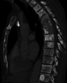 Sclerosis of the bones of the thoracic spine due to prostate cancer metastases (CT image)
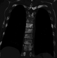 Sclerosis of the bones of the thoracic spine due to prostate cancer metastases (CT image)
Diagnosis
Osteosclerosis can be detected with a simple radiography. There are white portions of the bone which appear due to the increased number of bone trabeculae.
Animals
In the animal kingdom there also exists a non-pathological form of osteosclerosis, resulting in unusually solid bone structure with little to no marrow. It is often seen in aquatic vertebrates, especially those living in shallow waters,[7] providing ballast as an adaptation for an aquatic lifestyle. It makes bones heavier, but also more fragile. In those animal groups, osteosclerosis often occurs together with bone thickening (pachyostosis). This joint occurrence is called pachyosteosclerosis.
See also
- Pachyostosis
- Pachyosteosclerosis
- Heinrich Albers-Schönberg
References
- EL-Sobky TA, Elsobky E, Sadek I, Elsayed SM, Khattab MF (2016). "A case of infantile osteopetrosis: The radioclinical features with literature update". Bone Rep. 4:11-16. http://doi.org/10.1016/j.bonr.2015.11.002. PMC4926827. PMID 28326337
- Ihde LL, Forrester DM, Gottsegen CJ, Masih S, Patel DB, Vachon LA, et al. (2011). "Sclerosing bone dysplasias: Review and differentiation from other causes of osteosclerosis". RadioGraphics. 31:7, 1865-82. DOI: https://dx.doi.org/10.1148/rg.317115093
- Niederhauser, BD; Dingli, D; Kyle, RA; Ringler, MD (July 2014). "Imaging findings in 22 cases of Schnitzler syndrome: characteristic para-articular osteosclerosis, and the "hot knees" sign differential diagnosis". Skeletal Radiology. 43 (7): 905–15. doi:10.1007/s00256-014-1857-y. PMID 24652142.
- Soubrier, M; Dubost, JJ; Jouanel, P; Tridon, A; Flori, B; Leguille, C; Ristori, JM; Bussière, JL (1994). "[Multiple complications of monoclonal IgM]". La Revue de Médecine Interne. 15 (7): 484–6. doi:10.1016/s0248-8663(05)81474-2. PMID 7938961.
- Fiore CE, Riccobene S, Mangiafico R, Santoro F, Pennisi P (2005). "Hepatitis C-associated osteosclerosis (HCAO): report of a new case with involvement of the OPG/RANKL system" (PDF). Osteoporos Int. 16 (12): 2180–4. doi:10.1007/s00198-005-1858-8. PMID 15983730.
- EL-Sobky TA, El-Haddad A, Elsobky E, Elsayed SM, Sakr HM (2017). “Reversal of skeletal radiographic pathology in a case of malignant infantile osteopetrosis following hematopoietic stem cell transplantation”. Egypt J Radiol Nucl Med. 48 (1):237–43. http://doi.org/10.1016/j.ejrnm.2016.12.013.
- Houssaye, A. (2009). "Pachyostosis" in aquatic amniotes: a review. Integrative Zoology 4(4): 325-340.