Osteoid osteoma
An osteoid osteoma is a benign bone tumor that arises from osteoblasts and some components of osteoclasts and was originally thought to be a smaller version of an osteoblastoma. Osteoid osteomas tend to be less than 1.5 cm in size. The tumor can be in any bone in the body but are most common in long bones, such as the femur and tibia. They account for 10 to 12 percent of all benign bone tumors. "Osteoid osteomas may occur at any age, and are most common in patients between the ages of 4 and 25 years old. Males are affected approximately three times more commonly than females."[1]
| Osteoid osteoma | |
|---|---|
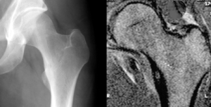 | |
| Osteoid osteoma of the trochanter minor: X-ray and MRI with marked sclerosis around the nidus | |
| Specialty | Oncology |
Signs and symptoms
The most common symptoms of an Osteoid Osteoma are:
- dull pain that escalates to severe at night OR slight pain, rising to become severe even at nighttime, affecting sleep quality
- limping
- muscle atrophy
- bowing deformity
- swelling
- increased or decreased bone growth
[2] [3] The most common symptom is pain that can be relieved with over the counter pain medication in the beginning. After the benign tumor develops further the pain can not be alleviated with medication and minor to severe swelling starts to occur. Although, in some cases the pain level remains the same for years, and regular NSAIDs intake keeps the pain at bay. The tumor is often found through x-ray imaging. "Conventional radiographs reveal a well-demarcated lytic lesion (nidus) surrounded by a distinct zone of sclerosis" that allow doctors to identify the tumor.[4]
Characterized by being less than 1.5 cm in diameter, osteoid osteomas most frequently occur in young men (Male:Female ratio 3:1) and may occur in any bone of the body, most frequently around the knee but often also seen in the vertebrae, in the long bones and less commonly in the mandible or other craniofacial bones.
Severe pain typically occurs at night, but can also be constant at any time of day.[5] The chief complaint may only be of dull pain which is non radiating and persistent throughout 24 hours but increases significantly at night. Pain tends to be relieved with NSAIDs such as ibuprofen.[6]
Histological findings
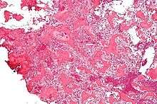
On histological examination, osteoid osteoma consists of a small (< 1.52 cm), yellowish-to-red nidus of osteoid and woven bone with interconnected trabeculae, and a background and rim of highly vascularized, fibrous connective tissue. Varying degrees of sclerotic bone reaction may surround the lesion. Benign osteoblastoma is virtually indistinguishable from osteoid osteoma. The usual appearance included a fibrovascular stroma with numerous osteoblasts, osteoid tissue, well-formed woven bone, and giant cells. The osteoblasts were generally small and regular in shape.[7]
Diagnosis
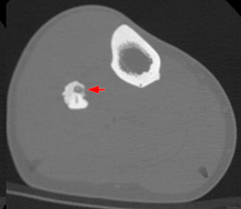
Radiographs in osteoid osteoma typically show a round lucency, containing a dense sclerotic central nidus (the characteristic lesion in this kind of tumor) surrounded by sclerotic bone. The nidus is seldom larger than 1.5 cm.
The lesion can in most cases be detected on CT scan, bone scans and angiograms. Plain radiographs are not always diagnostic. MRI adds little to the CT findings which are useful for localisation. Radionuclide scanning shows intense uptake which is useful for localisation at surgery using a hand held detector, and for confirmation that the entire lesion has been removed.[8][9]
Treatment
Pain may be relieved by nonsteroidal anti-inflammatory drugs.
Treatment varies based on the health of the patient. If he/she is otherwise healthy and is not significantly bothered by the pain, the tumor is treated symptomatically with anti-inflammatories. If this therapy fails or the location of the tumor could lead to growth disturbances, scoliosis, or osteoarthritis, surgical or percutaneous ablation may be considered.[10] If surgery is preferred, the individual may be referred to a podiatrist or an orthopedic surgeon to perform the procedure. Post-surgery therapy and strengthening may be needed, depending on the tumor location and health of the individual. While shown to be effective, surgical resection has many potential complications, including difficult intraoperative identification of the tumor, local recurrence from incomplete resection, and resection of weight bearing bone that can necessitate prolonged hospital stays and complicate recovery.[11]
Recently, CT guided radiofrequency ablation has emerged as a less invasive alternative to surgical resection. In this technique, which can be performed under conscious sedation, an RF probe is introduced into the tumor nidus through a cannulated needle under CT guidance and heat is applied locally to destroy tumor cells. Since the procedure was first introduced for the treatment of osteoid osteomas in the early 1990s,[12] it has been shown in numerous studies to be less invasive and expensive, to result in less bone destruction and to have equivalent safety and efficacy to surgical techniques, with 66 to 96% of patients reporting freedom from symptoms.[13][14][15] While initial success rates with RFA are high, symptom recurrence after RFA treatment has been reported, with some studies demonstrating a recurrence rate similar to that of surgical treatment.[16] As of July 17, 2014, treatment with incisionless surgery utilizing an MRI to guide high-intensity ultrasound waves to destroy a benign bone tumor in the leg has been demonstrated.[17]
References
- "Osteoid Osteoma - OrthoInfo - AAOS".
- "Osteoid Osteoma". Knol.
- "Bone and Soft Tissue Tumors: Benign Tumors". Dana-Farber Cancer Institute. Archived from the original on 2009-02-28. Retrieved 2011-04-25.
- Singh, Arun Pal. "Osteoid Osteoma-Diagnosis And Treatment". Archived from the original on 2011-04-26. Retrieved 2011-04-25.
- Mungo, David V.; Zhang, Xinping; O'Keefe, Regis J.; Rosier, Randy N.; Puzas, J. Edward; Schwarz, Edward M. (2002). "COX-1 and COX-2 expression in osteoid osteomas". Journal of Orthopaedic Research. 20 (1): 159–62. doi:10.1016/S0736-0266(01)00065-1. PMID 11853083.
- Atesok, Kivanc I.; Alman, Benjamin A.; Schemitsch, Emil H.; Peyser, Amos; Mankin, Henry (2011). "Osteoid osteoma and osteoblastoma". The Journal of the American Academy of Orthopaedic Surgeons. 19 (11): 678–89. doi:10.5435/00124635-201111000-00004. PMID 22052644.
- "Osteoid Osteomas and Osteoblastomas of the Spine".
- Osteoid Osteoma Imaging at eMedicine
- Lateur, L.; Baert, A. L. (1977). "Localisation and diagnosis of osteoid osteoma of the carpal area by angiography". Skeletal Radiology. 2 (2): 75–9. doi:10.1007/BF00360985.
- http://www.bonetumor.org/tumors-bone/osteoid-osteoma%5B%5D
- Sim, F. H.; Dahlin, C. D.; Beabout, J. W. (1975-03-01). "Osteoid-osteoma: diagnostic problems". J Bone Joint Surg Am. 57 (2): 154–159. doi:10.2106/00004623-197557020-00004. ISSN 0021-9355. PMID 1112841. Archived from the original on 2016-10-06. Retrieved 2016-08-07.
- Rosenthal, D I; Alexander, A; Rosenberg, A E; Springfield, D (1992-04-01). "Ablation of osteoid osteomas with a percutaneously placed electrode: a new procedure". Radiology. 183 (1): 29–33. doi:10.1148/radiology.183.1.1549690. ISSN 0033-8419. PMID 1549690.
- Weber, Marc-André; Sprengel, Simon David; Omlor, Georg W.; Lehner, Burkhard; Wiedenhöfer, Bernd; Kauczor, Hans-Ulrich; Rehnitz, Christoph (2015-04-25). "Clinical long-term outcome, technical success, and cost analysis of radiofrequency ablation for the treatment of osteoblastomas and spinal osteoid osteomas in comparison to open surgical resection". Skeletal Radiology. 44 (7): 981–993. doi:10.1007/s00256-015-2139-z. ISSN 0364-2348. PMID 25910709.
- Rosenthal, Daniel I.; Hornicek, Francis J.; Torriani, Martin; Gebhardt, Mark C.; Mankin, Henry J. (2003-10-01). "Osteoid Osteoma: Percutaneous Treatment with Radiofrequency Energy". Radiology. 229 (1): 171–175. doi:10.1148/radiol.2291021053. ISSN 0033-8419. PMID 12944597.
- Rimondi, Eugenio; Mavrogenis, Andreas F.; Rossi, Giuseppe; Ciminari, Rosanna; Malaguti, Cristina; Tranfaglia, Cristina; Vanel, Daniel; Ruggieri, Pietro (2011-08-14). "Radiofrequency ablation for non-spinal osteoid osteomas in 557 patients". European Radiology. 22 (1): 181–188. doi:10.1007/s00330-011-2240-1. ISSN 0938-7994. PMID 21842430.
- Rosenthal, Daniel I.; Hornicek, Francis J.; Wolfe, Michael W.; Jennings, L. Candace; Gebhardt, Mark C.; Mankin, Henry J. (1998-06-01). "Percutaneous Radiofrequency Coagulation of Osteoid Osteoma Compared with Operative Treatment*". J Bone Joint Surg Am. 80 (6): 815–21. CiteSeerX 10.1.1.1018.5024. doi:10.2106/00004623-199806000-00005. ISSN 0021-9355. PMID 9655099. Archived from the original on 2016-10-06. Retrieved 2016-08-07.
- Focused Ultrasound Foundation. "Bone tumor destroyed using incisionless surgery: First in North American child." ScienceDaily. ScienceDaily, 6 August 2014. <www.sciencedaily.com/releases/2014/08/140806142126.htm>.
External links
| Classification | |
|---|---|
| External resources |