Optic vesicle
The eyes begin to develop as a pair of diverticula from the lateral aspects of the forebrain. These diverticula make their appearance before the closure of the anterior end of the neural tube;[1][2] after the closure of the tube they are known as the optic vesicles. Previous studies of optic vesicles suggest that the surrounding extraocular tissues - the surface ectoderm and extraocular mesenchyme - are necessary for normal eye growth and differentiation.[3]
| Optic vesicle | |
|---|---|
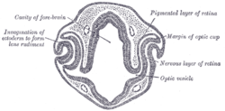 Transverse section of head of chick embryo of forty-eight hours’ incubation. (Optic vesicle labeled at lower right.) | |
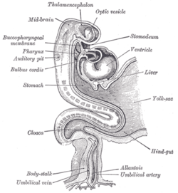 Human embryo about fifteen days old. Brain and heart represented from right side. Digestive tube and yolk sac in median section. (Optic vesicle labeled at center top.) | |
| Details | |
| Carnegie stage | 11 |
| Gives rise to | Human eyes |
| Identifiers | |
| Latin | vesicula optica; vesicula ophthalmica |
| TE | E5.14.3.4.2.2.4 |
| Anatomical terminology | |
They project toward the sides of the head, and the peripheral part of each expands to form a hollow bulb, while the proximal part remains narrow and constitutes the optic stalk.[4][5]
Additional images
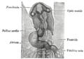 Head of chick embryo of about thirty-eight hours’ incubation, viewed from the ventral surface. X 26
References
This article incorporates text in the public domain from page 1001 of the 20th edition of Gray's Anatomy (1918)
Citations
- Hosseini, Hadi S.; Beebe, David C.; Taber, Larry A. (2014). "Mechanical effects of the surface ectoderm on optic vesicle morphogenesis in the chick embryo". Journal of Biomechanics. 47 (16): 3837–3846. doi:10.1016/j.jbiomech.2014.10.018. PMC 4261019. PMID 25458577.
- Hosseini, Hadi S.; Taber, Larry A. (2018). "How mechanical forces shape the developing eye". Progress in Biophysics and Molecular Biology. 137 (16): 25–36. doi:10.1016/j.pbiomolbio.2018.01.004. PMC 6085168. PMID 29432780.
- Fuhrmann, S. (2010). Eye Morphogenesis and Patterning of the Optic Vesicle. Current Topics in Developmental Biology Invertebrate and Vertebrate Eye Development, 61-84. doi:10.1016/b978-0-12-385044-7.00003-5
- Hosseini, Hadi S.; Beebe, David C.; Taber, Larry A. (2014). "Mechanical effects of the surface ectoderm on optic vesicle morphogenesis in the chick embryo". Journal of Biomechanics. 47 (16): 3837–3846. doi:10.1016/j.jbiomech.2014.10.018. PMC 4261019. PMID 25458577.
- Hosseini, Hadi S.; Taber, Larry A. (2018). "How mechanical forces shape the developing eye". Progress in Biophysics and Molecular Biology. 137 (16): 25–36. doi:10.1016/j.pbiomolbio.2018.01.004. PMC 6085168. PMID 29432780.
Sources
- Fuhrmann, S. (2010). Eye Morphogenesis and Patterning of the Optic Vesicle. Current Topics in Developmental Biology Invertebrate and Vertebrate Eye Development, 61-84. doi:10.1016/b978-0-12-385044-7.00003-5
External links
- eye-012—Embryo Images at University of North Carolina
- Overview at vision.ca
- Overview at temple.edu
| Authority control |
|
|---|