Ollier disease
Ollier disease is a rare nonhereditary sporadic disorder where intraosseous benign cartilaginous tumors (enchondroma) develop close to growth plate cartilage. Prevalence is estimated at around 1 in 100,000.[1]
| Ollier disease | |
|---|---|
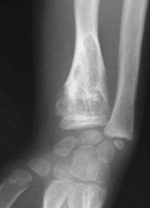 | |
| X-ray image showing enchondromas localized in the lower part of the radius of a 7-year-old girl with Ollier disease. | |
| Specialty | Medical genetics |
Presentation
Nominally, the disease consists of multiple enchondromas which usually develop in childhood. The growth of these enchondromas usually stops after skeletal maturation.[2] The affected extremity is shortened (asymmetric dwarfism) and sometimes bowed due to epiphyseal fusion anomalies. Persons with Ollier disease are prone to breaking bones and normally have swollen, aching limbs.
Associated conditions
Ollier disease carries a high risk of skeletal, visceral and brain malignancy which occurs in approximately 25% of patients. Juvenile granulosa cell tumour has been associated with the disease.[3] The incidence of secondary chondrosarcoma in Ollier disease is not known, but may be as high as 25%, pelvis and shoulder girdle being the commonest locations.[4] A related disorder called Maffucci syndrome named after Angelo Maffucci is characterized by enchondromas associated with multiple hemangiomas which usually occur in the hands and feet. Maffucci syndrome carries a higher risk for cancer.
Cause
Diagnosis
On radiographs, streaks of low density are seen projecting through the diaphyses into the epiphyses of the long bones, due to ectopic cartilage deposits. With age, the cartilage may calcify in the typical "snowflake" pattern.
Treatment
The deformities are managed surgically to preserve the function of the limb.[4]
Epidemiology
One person in every 100,000 is affected. Ollier disease is not normally diagnosed until toddler years because it is not very visible.
Additional images
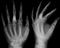 X-ray showing calcified enchondromas localized in finger a 37-year-old patient affected with Ollier disease
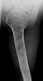 X-ray showing enchondromas localized in the humerus of a 37-year-old patient affected with Ollier disease
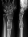 X-ray showing enchondromas localized in the lower part of the radius of a 37-year-old patient affected with Ollier disease
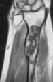 MRI showing enchondromas localized in the lower part of the radius of a 37-year-old patient affected with Ollier disease
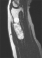 MRI showing enchondromas localized in the lower part of the radius of a 37-year-old patient affected with Ollier disease.
 Enchondromas localized in the upper part of the humerus of the same patient
See also
References
- Silve C, Jüppner H (2006). "Ollier disease". Orphanet J Rare Dis. 1: 37. doi:10.1186/1750-1172-1-37. PMC 1592482. PMID 16995932.
- Turek's orthopaedics principles and their application (6th ed.). Philadelphia: Lippincott Williams & Wilkins. 2005. p. 438. ISBN 9780781742986.
- Stephen C. Rubin (2001). Ovarian cancer (2nd ed.). Philadelphia [u.a.]: Lippincott Williams and Wilkins. ISBN 9780781724081.
- Lovell and Winter's pediatric orthopaedics (3rd ed.). Philadelphia: Lippincott. 1990. ISBN 9780397509140.
- synd/1813 at Who Named It?
External links
| Classification | |
|---|---|
| External resources |