Nipple adenoma
A nipple adenoma is a rare benign tumour of the breast.
| Nipple adenoma | |
|---|---|
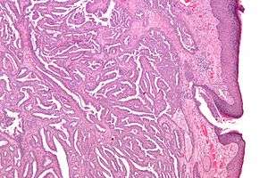 | |
| Micrograph of a nipple adenoma. H&E stain. | |
| Specialty | Oncology |
The condition may also be known as :
- Florid papillomatosis of the nipple
- Florid adenomatosis
- Subareolar duct papillomatosis
- Erosive adenomatosis[1]
Signs and symptoms
Nipple adenomas may be felt as a lump under the nipple or areola. They may come to attention because of nipple pain, ulceration, swelling or discharge.[1]
Diagnosis
Definition
A nipple adenoma is a type of intraductal papilloma that arises within the lactiferous ducts that are located within the nipple.[2]
Differential diagnosis
The microscopic appearance of a nipple adenoma can be mistaken for carcinoma.[1] Other conditions that have similar symptoms and signs as nipple adenoma include Paget's disease of the breast, other intraductal papillomas, ductal carcinoma in situ (DCIS), syringomatous adenoma of the nipple and subareolar sclerosing duct hyperplasia.[1]
Imaging
Lesions of the nipple and areola, such as nipple adenoma, may be difficult to image clearly on routine mammogram or ultrasonography. Nipple adenomas can be imaged using magnetic resonance imaging (MRI) and conventional or MR ductogram.[3]
Biopsy
Once excised, the macroscopic appearance of nipple adenomas is of a poorly defined nodular mass. The microscopic appearance can be quite bizarre, and may be misinterpreted as a carcinoma. Nipple adenomas usually have a rounded outline at low magnification, and at higher magnification can be seen to consist of a haphazardly arranged mass of proliferating tubular structures composed of epithelial and myoepithelial cells within varying amounts of fibrous stroma. The epithelial cells are usually columnar, but the columnar epithelial cells can undergo apocrine or squamous metaplasia. Mitotic figures and necrosis are not commonly seen.[1]
Treatment
The appropriate treatment in contemporary western medicine is complete surgical excision of the abnormal growth with a small amount of normal surrounding breast tissue.[1]
Prognosis
Nipple adenomas are non-cancerous growths, which can recur if not completely surgically removed.[1] There are reported cases of cancers arising within nipple adenomas, and following excision of nipple adenomas, but these are rare occurrences.[4]
Epidemiology
Nipple adenomas most commonly occur in 30- to 40-year-old women,[1] but can also occur in men.[5] They can also occur at any age, including in the elderly, in adolescence,[6] and in infants.[7]
References
- Stoler, Mark A.; Mills, Stacey E.; Carter, Darryl; Joel K Greenson; Reuter, Victor E. (2009). Sternberg's Diagnostic Surgical Pathology. Hagerstwon, MD: Lippincott Williams & Wilkins. ISBN 0-7817-7942-1.
- Pfeifer, John D.; Humphrey, Peter A.; Dehner, Louis P. (2008). The Washington Manual of surgical pathology. Philadelphia: Wolters Kluwer Health/Lippincott Williams & Wilkins. ISBN 0-7817-6527-7.
- Sarica O, Zeybek E, Ozturk E (July 2010). "Evaluation of nipple-areola complex with ultrasonography and magnetic resonance imaging". J Comput Assist Tomogr. 34 (4): 575–86. doi:10.1097/RCT.0b013e3181d74a88. PMID 20657228.
- Rao P, Shousha S (2010). "Male nipple adenoma with DCIS followed 9 years later by invasive carcinoma". Breast J. 16 (3): 317–8. doi:10.1111/j.1524-4741.2010.00900.x. PMID 20408826.
- Tuveri M, Calò PG, Mocci C, Nicolosi A (September 2010). "Florid papillomatosis of the male nipple". Am. J. Surg. 200 (3): e39–40. doi:10.1016/j.amjsurg.2009.10.026. PMID 20409515.
- Tao W, Kai F, Yue Hua L (2010). "Nipple adenoma in an adolescent". Pediatr Dermatol. 27 (4): 399–401. doi:10.1111/j.1525-1470.2010.01176.x. PMID 20653865.
- Clune JE, Kozakewich HP, VanBeek CA, Labow BI, Greene AK (November 2009). "Nipple adenoma in infancy". J. Pediatr. Surg. 44 (11): 2219–22. doi:10.1016/j.jpedsurg.2009.08.020. PMID 19944237.