Neutrophilia
Neutrophilia (also called neutrophil leukocytosis or occasionally neutrocytosis) is leukocytosis of neutrophils, that is, a high number of neutrophils in the blood.[1] Because neutrophils are the main type of granulocytes, mentions of granulocytosis often overlap in meaning with neutrophilia.
| Neutrophilia | |
|---|---|
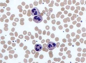 | |
| Neutrophils with a segmented nuclei surrounded by erythrocytes, the intra-cellular granules are visible in the cytoplasm (Giemsa stained) |
The opposite of neutrophilia is neutropenia.
Causes
Neutrophils are the primary white blood cells that respond to a bacterial infection, so the most common cause of neutrophilia is a bacterial infection, especially pyogenic infections.[2]
Neutrophils are also increased in any acute inflammation, so will be raised after a heart attack,[2] other infarct or burns.[2]
Some drugs, such as prednisone, have the same effect as cortisol and adrenaline (epinephrine), causing marginated neutrophils to enter the blood stream.
A neutrophilia might also be the result of a malignancy. Chronic myelogenous leukemia (CML or chronic myeloid leukaemia) is a disease where the blood cells proliferate out of control. These cells may be neutrophils. Neutrophilia can also be caused by appendicitis and splenectomy.[3]
Primary neutrophilia can additionally be a result of leukocyte adhesion deficiency.[4]
"Left shift"
A "left shift" refers to the presence of increased proportions of younger, less well differentiated neutrophils and neutrophil-precursor cells in the blood. This generally reflects early or premature release of myeloid cells from the bone marrow, the site where neutrophils are generated. A severe neutrophilia with left shift is referred to as a leukemoid reaction. The leukocyte alkaline phosphatase (LAP) score, which refers to the amount of alkaline phosphatase per neutrophil, will increase. In a severe infection, toxic granulation changes happen to the neutrophils.
This can resemble Pelger-Huet anomaly.[5][6]
See also
References
- "neutrophilia" at Dorland's Medical Dictionary
- Table 12-6 in: Mitchell, Richard Sheppard; Kumar, Vinay; Abbas, Abul K; Fausto, Nelson. Robbins Basic Pathology. Philadelphia: Saunders. ISBN 1-4160-2973-7. 8th edition.
- "bloodandcancerclinic". Archived from the original on 21 May 2013. Retrieved 10 April 2013.
- "Titre" (PDF). Retrieved 2019-11-16.
- Mohamed IS, Wynn RJ, Cominsky K, et al. (June 2006). "White blood cell left shift in a neonate: a case of mistaken identity". J Perinatol. 26 (6): 378–80. doi:10.1038/sj.jp.7211513. PMID 16724080.
- Shmuely H, Pitlik SD, Inbal A, Rosenfeld JB (June 1993). "Pelger-Huët anomaly mimicking 'shift to the left'". Neth J Med. 42 (5–6): 168–70. PMID 8377874.
External links
| Classification | |
|---|---|
| External resources |