Neuropeptide
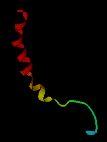
Neuropeptides are small protein-like molecules (peptides) used by neurons to communicate with each other. They are neuronal signalling molecules that influence the activity of the brain and the body in specific ways. Different neuropeptides are involved in a wide range of brain functions, including analgesia, reward, food intake, metabolism, reproduction, social behaviors, learning and memory.

Mechanism and Synthesis
Neuropeptides are synthesized from large, inactive precursor proteins called prepropeptides, which are cleaved into several active peptides. Prepropeptides can contain multiple copies of the same peptide, as well as sequences for different peptides, depending on the organism. The human genome contains about 90 genes that encode precursors of neuropeptides. At present about 100 different peptides are known to be released by different populations of neurons in the mammalian brain.[1] In Drosophila, there are 50 known genes that encode neuropeptides. These peptides are synthesized at the soma, entered into the secretory pathway to pass through the rER-Golgi complex, further processed, then packaged into large dense-core vesicles for transport down the axon or dendrites.[2][3] Neuropeptides are released in a calcium-dependent manner to bind to G-protein coupled receptors (GPCR). The relationship between neuropeptide and corresponding GPCRs are highly conserved and specific. There are as many genes encoding GPCRs in Drosophila as there are neuropeptides .[2][3] Large dense core vesicles release low volumes of neuropeptide compared to synaptic vesicles and neurotransmitters. Neuropeptides are not immediately reuptaken, degraded or recycled and thus are bioactive for long periods of time.[2]
Peptidergic expression in the brain can be highly selective and specific. In Drosophila larvae for example, eclosion hormone is expressed in just two neurons and SIFamide is expressed in four.[3] In contrast to its selective expression, peptidergic activity can be broad and long-lasting. Peptides are not recycled back into the cell once secreted, unlike conventional neurotransmitters (glutamate, dopamine, serotonin). Thus, neuropeptides can diffuse across tissues and are bioactive for long periods of time.[2]
There is a vast array of bioactive peptides which yields a diversity of effects. Peptidergic neurons commonly express and synthesize multiple neuropeptides and also release other small-molecule neurotransmitters, but the co-release relationships are poorly understood.[4] This diversity is in part due to tissue-specific processing of neuropeptide precursors. Different tissues have tailored post-translational processing steps which yield structurally and functionally different peptides.[2]
Comparison to Neurotransmitters
Generally, peptides act at metabotropic or G-protein-coupled receptors expressed by selective populations of neurons. In essence they act as specific signals between one population of neurons and another. Neurotransmitters generally affect the excitability of other neurons, by depolarising them or by hyperpolarising them. Peptides have much more diverse effects; amongst other things, they can affect gene expression, local blood flow, synaptogenesis, and glial cell morphology. Peptides tend to have prolonged actions, and some have striking effects on behaviour.
Neurons very often make both a conventional neurotransmitter (such as glutamate, GABA or dopamine) and one or more neuropeptides. Peptides are generally packaged in large dense-core vesicles, and the co-existing neurotransmitters in small synaptic vesicles. The large dense-core vesicles are often found in all parts of a neuron, including the soma, dendrites, axonal swellings (varicosities) and nerve endings, whereas the small synaptic vesicles are mainly found in clusters at presynaptic locations.[5][6] Release of the large vesicles and the small vesicles is regulated differently.
Examples
Many populations of neurons have distinctive biochemical phenotypes. For example, in one subpopulation of about 3000 neurons in the arcuate nucleus of the hypothalamus, three anorectic peptides are co-expressed: α-melanocyte-stimulating hormone (α-MSH), galanin-like peptide, and cocaine-and-amphetamine-regulated transcript (CART), and in another subpopulation two orexigenic peptides are co-expressed, neuropeptide Y and agouti-related peptide (AGRP). These are not the only peptides in the arcuate nucleus; β-endorphin, dynorphin, enkephalin, galanin, ghrelin, growth-hormone releasing hormone, neurotensin, neuromedin U, and somatostatin are also expressed in subpopulations of arcuate neurons. These peptides are all released centrally and act on other neurons at specific receptors. The neuropeptide Y neurons also make the classical inhibitory neurotransmitter GABA.
Invertebrates also have many neuropeptides. CCAP has several functions including regulating heart rate, allatostatin and proctolin regulate food intake and growth, bursicon controls tanning of the cuticle and corazonin has a role in cuticle pigmentation and moulting.
Peptide signals play a role in information processing that is different from that of conventional neurotransmitters, and many appear to be particularly associated with specific behaviours. For example, oxytocin and vasopressin have striking and specific effects on social behaviours, including maternal behaviour and pair bonding. The following is a list of neuroactive peptides coexisting with other neurotransmitters. Transmitter names are shown in bold.
Norepinephrine (noradrenaline). In neurons of the A2 cell group in the nucleus of the solitary tract), norepinephrine co-exists with:
- Galanin
- Enkephalin
- Neuropeptide Y
GABA
- Somatostatin (in the hippocampus)
- Cholecystokinin
- Neuropeptide Y (in the arcuate nucleus)
- VIP
- Substance P
- Cholecystokinin
- Neurotensin
- Glucagon-like peptide-1 (in the nucleus accumbens)
Epinephrine (adrenaline)
- Neuropeptide Y
- Neurotensin
Serotonin (5-HT)
- Substance P
- TRH
- Enkephalin
Some neurons make several different peptides. For instance, Vasopressin co-exists with dynorphin and galanin in magnocellular neurons of the supraoptic nucleus and paraventricular nucleus, and with CRF (in parvocellular neurons of the paraventricular nucleus)
Oxytocin in the supraoptic nucleus co-exists with enkephalin, dynorphin, cocaine-and amphetamine regulated transcript (CART) and cholecystokinin.
References
- database of all neuropetides
- Mains, Richard E.; Eipper, Betty A. (1999). "The Neuropeptides". Basic Neurochemistry: Molecular, Cellular and Medical Aspects. 6th edition.
- Nässel, Dick R.; Zandawala, Meet (August 2019). "Recent advances in neuropeptide signaling in Drosophila, from genes to physiology and behavior". Progress in Neurobiology. 179: 101607. doi:10.1016/j.pneurobio.2019.02.003. ISSN 1873-5118. PMID 30905728.
- Nässel, Dick R. (23 March 2018). "Substrates for Neuronal Cotransmission With Neuropeptides and Small Molecule Neurotransmitters in Drosophila". Frontiers in Cellular Neuroscience. 12. doi:10.3389/fncel.2018.00083. ISSN 1662-5102. PMC 5885757. PMID 29651236.
- van den Pol AN (October 2012). "Neuropeptide transmission in brain circuits". Neuron. 76 (1): 98–115. doi:10.1016/j.neuron.2012.09.014. PMC 3918222. PMID 23040809.
- Leng G, Ludwig M (December 2008). "Neurotransmitters and peptides: whispered secrets and public announcements". The Journal of Physiology. 586 (23): 5625–32. doi:10.1113/jphysiol.2008.159103. PMC 2655398. PMID 18845614.
| Wikimedia Commons has media related to Neuropeptide. |
External links
| Look up neuropeptide in Wiktionary, the free dictionary. |
- Neuropeptides Journal
- Neuropeptides reference website (a comprehensive neuropeptide database)
- Neuropeptides eBook series
- Neuropeptide chapter in the C. elegans Wormbook excellent, and very accessible, discussion of neuropeptide biology in C. elegans