Neurogenic bladder dysfunction
Neurogenic bladder dysfunction, or neurogenic bladder, refers to urinary bladder problems due to disease or injury of the central nervous system or peripheral nerves involved in the control of urination. There are multiple types of neurogenic bladder depending on the underlying cause and the symptoms. Symptoms include overactive bladder, urinary urgency, frequency, incontinence or difficulty passing urine. A range of diseases or conditions can cause neurogenic bladder including spinal cord injury, multiple sclerosis, stroke, brain injury, spina bifida, peripheral nerve damage, Parkinson's disease, or other neurodegenerative diseases. Neurogenic bladder can be diagnosed through a history and physical as well as imaging and more specialized testing. Treatment depends on underlying disease as well as symptoms and can be managed with behavioral changes, medications, surgeries, or other procedures. The symptoms of neurogenic bladder, especially incontinence, can have a significant impact on quality of life.
Classification
There are different types of neurogenic bladder depending on the underlying cause. Many of these types may have similar symptoms.
Uninhibited
Uninhibited bladder is usually due to damage to the brain from a stroke or brain tumor. This can cause reduced sensation of bladder fullness, low capacity bladder and urinary incontinence. Unlike other forms of neurogenic bladder, it does not lead to high bladder pressures that can cause kidney damage.[1]
Spastic
In spastic neurogenic bladder (also known as upper motor neuron or hyper-reflexive bladder), the muscle of the bladder (detrusor) and urethral sphincter do not work together and are usually tightly contracted at the same time. This phenomenon is also called detrusor external sphincter dyssynergia (DESD). This leads to urinary retention with high pressures in the bladder that can damage the kidneys. The bladder volume is usually smaller than normal due to increased muscle tone in the bladder. Spastic neurogenic bladder is usually caused by damage to the spinal cord above the level of the 10th thoracic vertebrae (T10).[1][2]
Flaccid
In flaccid bladder (also known as lower motor neuron or hypotonic bladder), the muscles of the bladder lose ability to contract normally. This can cause the inability to void urine even if the bladder is full and cause a large bladder capacity. The internal urinary sphincter can contract normally, however urinary incontinence is common. This type of neurogenic bladder is caused by damage to the peripheral nerves that travel from the spinal cord to the bladder.[1]
Mixed
Mixed type of neurogenic bladder can cause a combination of the above presentations. In mixed type A, the bladder muscle is flaccid but the sphincter is overactive. This creates a large, low pressure bladder and inability to void, but does not carry as much risk for kidney damage as a spastic bladder. Mixed type B is characterized by a flaccid external sphincter and a spastic bladder causing problems with incontinence.[1]
Signs and symptoms
Neurogenic bladder can cause a range of urinary symptoms including urinary urgency, urinary incontinence or difficulty urinating (urinary retention.) The first sign of bladder dysfunction may be recurrent urinary tract infections (UTIs).
Causes
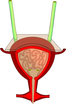
Urine storage and elimination (urination) requires coordination between the bladder emptying muscle (detrusor) and the external sphincter of the bladder. This coordination can be disrupted by damage or diseases of the central nervous system, peripheral nerves or autonomic nervous system.[3] This includes any condition that impairs bladder signaling at any point along the path from the urination center in the brain, spinal cord, peripheral nerves and the bladder.
Central nervous system
Damage to the brain or spinal cord is the most common cause of neurogenic bladder. Damage to the brain can be caused by stroke, brain tumors, multiple sclerosis, Parkinson's disease or other neurodegenerative conditions.[3] Bladder involvement is more likely if the damage is in the area of the pons. Damage to the spinal cord can be caused by traumatic injury, demyelinating disease, syringomyelia, cauda equina syndrome, or spina bifida. Spinal cord compression from herniated disks, tumor, or spinal stenosis can also result in neurogenic bladder.[1][3]
Peripheral nervous system
Damage to the nerves that travel from the spinal cord to the bladder (peripheral nerves) can cause neurogenic bladder, usually the flaccid type. Nerve damage can be caused by diabetes, alcoholism, and vitamin B12 deficiency. Peripheral nerves can also be damaged as a complication of major surgery of the pelvis, such as for removal of tumors.[1]
Diagnosis
The diagnosis of neurogenic bladder is made based on a complete history and physical examination and may require imaging and specialized studies. History should include information on the onset, duration, triggers, severity, other medical conditions and medications (including anticholinergics, calcium channel blockers, diuretics, sedatives, alpha-adrenergic agonist, alpha 1 antagonists).[2][3] Urinary symptoms may include frequency, urgency, incontinence or recurrent urinary tract infections (UTIs). Questionnaires can be helpful in quantifying symptom burden.[2] In children it is important to obtain a prenatal and developmental history.[4]
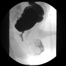
Ultrasound imaging can give information on the shape of the bladder, post-void residual volume, and evidence of kidney damage such as kidney size, thickness or ureteral dilation. A voiding cystourethrography study uses contrast dye to obtain images of the bladder both when it is full and after urination which can show changes in bladder shape consistent with neurogenic bladder.[4]
Urodynamic studies are an important component of the evaluation for neurogenic bladder. Urodynamics refers to the measurement of the pressure-volume relationship in the bladder. The bladder usually stores urine at low pressure and urination can be completed without a dramatic pressure rise. Damage to the kidneys is probable if the pressure rises above 40 cm of water during filling.[2] Bladder pressure can be measured by cystometry, during which the bladder is artificially filled with a catheter and bladder pressures and detrusor activity are monitored. Patterns of involuntary detrusor activity as well as bladder flexibility, or compliance, can be evaluated. The most valuable test to test for detrusor sphincter dyssynergia (DESD) is to perform cystometry simultaneously with external sphincter electromyography (EMG).[3] Uroflowmetry is a less-invasive study that can measure urine flow rate and use it to estimate detrusor strength and sphincter resistance.[2][5] Urethral pressure monitoring is another less-invasive approach to assessing detrusor sphincter dyssynergia.[5] These studies can be repeated at regular intervals, especially if symptoms worsen or to measure response to therapies.[4]
Evaluation of kidney function through blood tests such as serum creatinine should be obtained.[2]
Imaging of the pelvis with CT scan or magnetic resonance imaging may be necessary, especially if there is concern for an obstruction such as a tumor. The inside of the bladder can be visualized by cystoscopy.
Treatment
Treatment depends on the type of neurogenic bladder and other medical problems. Treatment strategies include catheterization, medications, surgeries or other procedures. The goals of treatment is to keep bladder pressures in a safe range and eliminate residual urine in the bladder after urination (post-void residual volumes).
Catherization
Emptying the bladder with the use of a catheter is the most common strategy for managing urinary retention from neurogenic bladder. For most patients, this can be accomplished with intermittent catherization which involves no surgery or permanently attached appliances. Intermittent catheterization involves using straight catheters (which are usually disposable or single-use products) several times a day to empty the bladder.[3] This can be done independently or with assistance. For people who are unable to use disposable straight catheters, a Foley catheter allows continuous drainage of urine into a sterile drainage bag that is worn by the patient but are associated with higher rates of complications.[6]
Medications
Oxybutynin is a common anti-cholinergic medication used to reduce bladder contractions by blocking M3 muscarinic receptors in the detrusor.[6] Its use is limited by side effects such as dry mouth, constipation and decreased sweating. Tolterodine is a longer acting anticholinergic that may have fewer side effects.[4]
For urinary retention, cholinergics (muscarinic agonists) like bethanechol can improve the squeezing ability of the bladder. Alpha blockers can also reduce outlet resistance and allow complete emptying if there is adequate bladder muscle function.[4]
Botulinum Toxin
Botulinum toxin (Botox) can be used through two different approaches. For spastic neurogenic bladder, the bladder muscle (detrusor) can be injected which will cause it to be flaccid for 6-9 months. This prevents high bladder pressures and intermittent catherization must be used during this time.[4]
Botox can also be injected into the external sphincter to paralyze a spastic sphincter in patients with detrusor sphincter dyssynergia.[5]
Neuromodulation
There are various strategies to alter the interaction between the nerves and muscles of the bladder, including nonsurgical therapies (transurethral electrical bladder stimulation), minimally invasive procedures (sacral neuromodulation pacemaker), and operative (reconfiguration of sacral nerve root anatomy).[4]
Surgery
Surgical interventions may be pursued if medical approaches have been maximized. Surgical options include creation of a stoma that bypasses the urethra to empty the bladder directly. This conduit may be created from the appendix (Mitrofanoff stoma) or a portion of the ileum (‘Yang-Monti’ conduit).[4] The ileum and [[ascending colon can also be used to create a pouch accessible for catherization (Indiana pouch). Urethral stents or urethral sphincterotomy are other surgical approaches that can reduce bladder pressures but require use of an external urinary collection device.[5]
Epidemiology
The overall prevalence of neurogenic bladder is limited due to the broad range of conditions that can lead to urinary dysfunction. Neurogenic bladder is common with spinal cord injury and multiple sclerosis.[5] Rates of some type of urinary dysfunction surpass 80% one year after spinal cord injury.[6] Among patients with multiple sclerosis, 20–25% will develop neurogenic bladder although the type and severity bladder dysfunction is variable.[5]
Complications
Neurogenic bladder can cause hydronephrosis, recurrent urinary tract infections, and recurrent kidney stones which may compromise kidney function.[6] This is especially significant in spastic neurogenic bladder that leads to high bladder pressures. Kidney failure was previously a leading cause of mortality in patients with spinal cord injury but is now dramatically less common due to improvements in bladder management.[6]
References
- Dorsher PT, McIntosh PM (2012). "Neurogenic bladder". Advances in Urology. 2012: 816274. doi:10.1155/2012/816274. PMC 3287034. PMID 22400020.
- Amarenco, Gerard; Sheikh Ismaël, Samer; Chesnel, Camille; Charlanes, Audrey; LE Breton, Frederique (Dec 2017). "Diagnosis and clinical evaluation of neurogenic bladder". European Journal of Physical and Rehabilitation Medicine. 53 (6): 975–980. doi:10.23736/S1973-9087.17.04992-9. ISSN 1973-9095. PMID 29072046.
- Bacsu, Chasta-Dawne; Chan, Lewis; Tse, Vincent (2012). "Diagnosing detrusor sphincter dyssynergia in the neurological patient". BJU International. 109 Suppl 3: 31–34. doi:10.1111/j.1464-410X.2012.11042.x. ISSN 1464-410X. PMID 22458490.
- Sripathi, Venkataramani; Mitra, Aparajita (2017-07-01). "Management of Neurogenic Bladder". The Indian Journal of Pediatrics. 84 (7): 545–554. doi:10.1007/s12098-017-2356-7. ISSN 0973-7693. PMID 28553689.
- Stoffel, John T. (2016). "Detrusor sphincter dyssynergia: a review of physiology, diagnosis, and treatment strategies". Translational Andrology and Urology. 5 (1): 127–135. doi:10.3978/j.issn.2223-4683.2016.01.08. ISSN 2223-4691. PMC 4739973. PMID 26904418.
- Schurch, Brigitte; Tawadros, Cécile; Carda, Stefano (2015). "Dysfunction of lower urinary tract in patients with spinal cord injury". Handbook of Clinical Neurology. 130: 247–267. doi:10.1016/B978-0-444-63247-0.00014-6. ISBN 9780444632470. ISSN 0072-9752. PMID 26003248.
External links
| Classification | |
|---|---|
| External resources |