Nasociliary nerve
The nasociliary nerve is a branch of the ophthalmic nerve (CN V1; one of three branches of the trigeminal nerve a.k.a. CN V). It is intermediate in size between the two other main branches of the ophthalmic nerve, the frontal nerve and the lacrimal nerve, and is more deeply placed.
| Nasociliary nerve | |
|---|---|
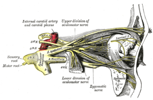 Nerves of the orbit, and the ciliary ganglion. Side view. (Nasociliary is at center.) | |
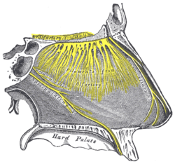 Nerves of septum of nose. Right side. (Nasociliary is rightmost yellow line.) | |
| Details | |
| From | Ophthalmic nerve |
| To | long root of the ciliary ganglion, the long ciliary nerves, the infratrochlear nerve, and the ethmoidal nerves |
| Identifiers | |
| Latin | nervus nasociliaris |
| TA | A14.2.01.025 |
| FMA | 52668 |
| Anatomical terms of neuroanatomy | |
Structure
The nasociliary nerve enters the orbit between the two heads of the lateral rectus muscles and between the superior and inferior rami of the oculomotor nerve (CN III). It passes across the optic nerve (CN II) and runs obliquely beneath the superior rectus muscle and superior oblique muscle to the medial wall of the orbital cavity. It passes through the anterior ethmoidal opening as the anterior ethmoidal nerve and enters the cranial cavity just below the cribriform plate of the ethmoid bone. It supplies branches to the mucous membrane of the nasal cavity and finally emerges between the inferior border of the nasal bone and the side nasal cartilages as the external nasal branch.
Function
A branch of the ophthalmic nerve [CN V1] in the superior orbital fissure, passing through the orbit, giving rise to the communicating branch to the ciliary ganglion, the long ciliary nerves, the posterior and anterior ethmoidal nerves, and terminating as the infratrochlear and nasal branches, which supply the mucous membrane of the nose, the skin of the tip of the nose, and the conjunctiva.[1]
Branches
The nasociliary nerve gives off the following branches:
- ethmoidal nerves
- infratrochlear nerve
- long ciliary nerve
- communicating branch to the ciliary ganglion (long root of the ciliary ganglion)
PLICA is a mnemonic often used to remember these branches.
Clinical significance
Testing
Since both the short and long ciliary nerves carry the afferent limb of the corneal blink reflex, one can test the integrity of the nasociliary nerve (and, ultimately, the trigeminal nerve) by examining this reflex in the patient. Normally both eyes should blink when either cornea (not the conjunctiva, which is supplied by the adjacent cutaneous nerves) is irritated. If neither eye blinks, then either the ipsilateral nasociliary nerve is damaged, or the facial nerve (CN VII, which carries the efferent limb of this reflex) is bilaterally damaged. If only the contralateral eye blinks, then the ipsilateral facial nerve is damaged. If only the ipsilateral eye blinks, then the contralateral facial nerve is damaged.
Additional images
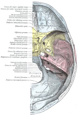 Base of the skull. Upper surface.
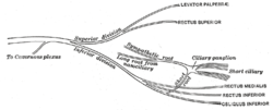 Plan of oculomotor nerve.
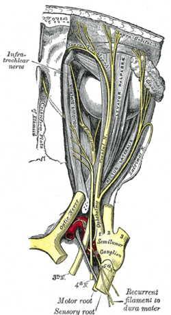 Nerves of the orbit. Seen from above.
 Distribution of the maxillary and mandibular nerves, and the submaxillary ganglion.
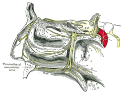 The sphenopalatine ganglion and its branches.
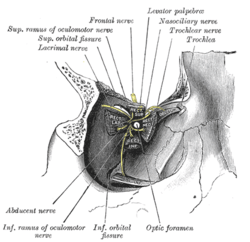 Dissection showing origins of right ocular muscles, and nerves entering by the superior orbital fissure.
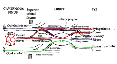 Pathways in the ciliary ganglion.
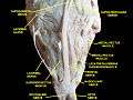 Extrinsic eye muscle. Nerves of orbita. Deep dissection.
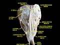 Extrinsic eye muscle. Nerves of orbita. Deep dissection.
References
This article incorporates text in the public domain from page 888 of the 20th edition of Gray's Anatomy (1918)
External links
- Anatomy figure: 29:03-08 at Human Anatomy Online, SUNY Downstate Medical Center - "A deeper dissection of the right orbit from a superior approach."
- lesson9 at The Anatomy Lesson by Wesley Norman (Georgetown University) (nasalseptumner)
- cranialnerves at The Anatomy Lesson by Wesley Norman (Georgetown University) (V)
- MedEd at Loyola GrossAnatomy/h_n/cn/cn1/cnb1.htm
| Authority control |
|---|