Myelography
Myelography is a type of radiographic examination that uses a contrast medium to detect pathology of the spinal cord, including the location of a spinal cord injury, cysts, and tumors. Historically the procedure involved the injection of a radiocontrast agent into the cervical or lumbar spine, followed by several X-ray projections. Today, myelography has largely been replaced by the use of MRI scans, although the technique is still sometimes used under certain circumstances – though now usually in conjunction with CT rather than X-ray projections.
| Myelography | |
|---|---|
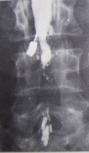 Myelogram showing arachnoiditis in the lumbar spine. | |
| MeSH | D009192 |
| OPS-301 code | 3-130 |
Uses
A myelogram is sometimes used to better image the spinal cord in patients with lumbar spinal stenosis.[1]
Procedure
A CT scan is typically performed after radiographic contrast media (dye) has been placed with fluoroscopic guidance into a sac-like lining (the first- and hardest and outermost- layer of the spinal meninges, the spinal dura mater) surrounding the spinal cord and nerves. The material is typically water-soluble, which has largely replaced nonsoluble oil-based fluids, while CT has largely replaced the conventional X-ray projections used for image acquisition in the past.
The process usually involves lying face down on a table, with the lower extremities secured tightly with straps to the table. After the skin area has been numbed, the dye is injected into the thecal sac, then the table is slowly rotated in a circular motion, first down at the head end for approximately 4 to 6 minutes, then rotated up at the head end for the same duration. Several more minutes lying flat and the process is complete. This movement ensures the contrast has sufficiently worked its way through the spinal cord, followed by X-rays or a CT scan.
Post-procedure care centers around ensuring that infection (especially skin or subcutaneous infections, myelitis or meningitis or encephalitis, or sepsis) does not set in and that the "plug" at the site of the spinal tap does not become dislodged. Patients are usually instructed to avoid strenuous activity and heavy lifting, for example. Some patients are given instructions to keep their heads elevated at least 30 degrees for a specified number of hours. Complications from the surgery can cause a loss of cerebrospinal fluid (CSF), which could cause severe headaches. This can be corrected by returning to the medical facility and having them perform a blood patch. In this procedure, a small amount of blood is taken from the arm and injected into the exact spinal tap location to stop the leaking of CSF.
Decline in use due to MRI
Nowadays, MRI is used for most investigations previously performed by myelography.[2] This has many advantages and does not require contrast fluid to be injected into the spine. However, a CT myelogram may be useful for patients who cannot undergo MRI (e.g., those with pacemakers or cochlear implants), or for those in whom MRI provides limited information (e.g., those with extensive metal in the spine).
Contrast agent
Prior to the late 1970s, iofendylate (trade names: Pantopaque, Myodil) was the radiocontrast agent typically employed in the procedure. It was an iodinated oil-based substance that the physician performing the spinal tap usually attempted to remove at the end of the procedure. This step was both difficult and painful and complete removal could not always be achieved. The process of removing the contrast agent necessitated removing some of the patient's CSF along with it and the resulting deficiency of CSF gave rise to severe headache if the patient was raised from the prone position, requiring bed rest in the laying position. Moreover, iofendylate's persistence in the body might sometimes lead to arachnoiditis, a potentially painful and debilitating lifelong disorder of the spine.[3][4] This led to extensive litigation around the world since the substance was administered to millions of myelography patients over the course of more than three decades.[5] After water-soluble agents (such as metrizamide) became available it was no longer necessary to remove the contrast agent as it would eventually be absorbed into the body although the water-soluble agent sometimes gave rise to severe headaches if it got into the head, requiring bed rest in the upright position.
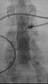 Myelography punction
 Conventional myelography in oblique projection. You can see the individual nerve root sheaths.
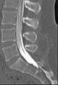 Computed tomography after conventional myelography. The overlap-free representation often allows a more secure assessment. The high density of contrast material may be troublesome in case of insufficient mixing prior to CT.
References
- Katz, Jeffrey N.; Harris, Mitchel B. (February 21, 2008). "Lumbar Spinal Stenosis". New England Journal of Medicine. 358 (8): 818–825. doi:10.1056/NEJMcp0708097. PMID 18287604.
- Leeds, NE; Kieffer, SA (November 2000). "Evolution of diagnostic neuroradiology from 1904 to 1999" (PDF). Radiology. 217 (2): 309–18. doi:10.1148/radiology.217.2.r00nv45309. PMID 11058623.
- Dunlevy, Sue (10 December 2016). "Australians crippled and in chronic pain from dye used in toxic X-rays". The Daily Telegraph. Retrieved 27 October 2017.
- William P. Dillon; Christopher F. Dowd (2014). "Chapter 53 – Neurologic Complications of Imaging Procedures". Aminoff's Neurology and General Medicine (5th ed.). Elsevier Academic Press. pp. 1089–1105. ISBN 978-0-12-407710-2.
- Myodil litigation
- Bontranger, Kenneth L. & Lampignano, John P. (2005). Radiographic Positioning and Related Anatomy, St. Louis: Elsevier Mosby. ISBN 0-323-02507-2.
External links
| X-ray/ Radiography |
| ||||||||||||
|---|---|---|---|---|---|---|---|---|---|---|---|---|---|
| MRI | |||||||||||||
| Ultrasound | |||||||||||||
| Radionuclide |
| ||||||||||||
| Optical/Laser | |||||||||||||
| Thermography |
| ||||||||||||
| Target conditions |
| ||||||||||||
| |||||||||||||