Muscles of mastication
There are four classical muscles of mastication. During mastication, three muscles of mastication (musculi masticatorii) are responsible for adduction of the jaw, and one (the lateral pterygoid) helps to abduct it. All four move the jaw laterally. Other muscles, usually associated with the hyoid, such as the mylohyoid muscle, are responsible for opening the jaw in addition to the lateral pterygoid.
| Muscles of mastication | |
|---|---|
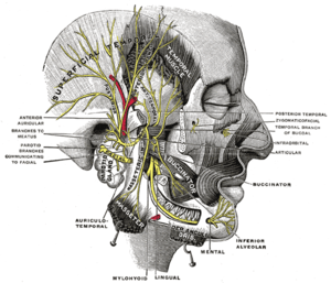 Mandibular division of the trigeminal nerve. | |
| Details | |
| Nerve | mandibular nerve |
| Identifiers | |
| Latin | musculi masticatorii |
| MeSH | D008410 |
| TA | A04.1.04.001 |
| FMA | 71437 74060, 71437 |
| Anatomical terms of muscle | |
Structure
The muscles are:
- The masseter (composed of the superficial and deep head)
- The temporalis (the sphenomandibularis is considered a part of the temporalis by some sources, and a distinct muscle by others)
- The medial pterygoid
- The lateral pterygoid
In humans, the mandible, or lower jaw, is connected to the temporal bone of the skull via the temporomandibular joint. This is an extremely complex joint which permits movement in all planes. The muscles of mastication originate on the skull and insert into the mandible, thereby allowing for jaw movements during contraction.
Each of these primary muscles of mastication is paired, with each side of the mandible possessing one of the four.
Innervation
Unlike most of the other facial muscles, which are innervated by the facial nerve (or CN VII), the muscles of mastication are innervated by the trigeminal nerve (or CN V). More specifically, they are innervated by the mandibular branch, or V3.
Development
This is a testament to their shared embryological origin from the first pharyngeal arch.
The muscles of facial expression, on the other hand, derive from the second pharyngeal arch.
Function
The mandible is the only bone that moves during mastication and other activities, such as talking.
While these four muscles are the primary participants in mastication, other muscles are usually if not always helping the process, such as those of the tongue and the cheeks.
Clinical significance
External links
| Wikimedia Commons has media related to Muscles of mastication. |
- Masticatory+Muscles at the US National Library of Medicine Medical Subject Headings (MeSH)
- http://www.med.umich.edu/lrc/coursepages/m1/anatomy2010/html/nervous_system/infratemp_lecture.html
| Authority control |
|---|