Morula
A morula (Latin, morus: mulberry) is an early-stage embryo consisting of 16 cells (called blastomeres) in a solid ball contained within the zona pellucida.[1][2]
| Morula | |
|---|---|
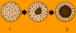 Blastulation. 1 - morula, 2 - blastula. | |
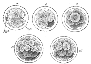 First stages of segmentation of a fertilized mammalian ovum. Semidiagrammatic. z.p. Zona pellucida. p.gl. Polar bodies. a. Two-cell stage. b. Four-cell stage. c. Eight-cell stage. d, e. Morula stage. | |
| Details | |
| Days | 3 |
| Precursor | Zygote |
| Gives rise to | Blastula, Blastocyst |
| Identifiers | |
| MeSH | D009028 |
| TE | E2.0.1.2.0.0.11 |
| Anatomical terminology | |
A morula is distinct from a blastocyst in that a morula (3–4 days after fertilization) is a mass of 16 totipotent cells in a spherical shape whereas a blastocyst (4–5 days after fertilization) has a cavity inside the zona pellucida along with an inner cell mass. A morula, if untouched and allowed to remain implanted, will eventually develop into a blastocyst.[3]
The morula is produced by a series of cleavage divisions of the early embryo, starting with the single-celled zygote. Once the embryo has divided into 16 cells, it begins to resemble a mulberry, hence the name morula (Latin, morus: mulberry).[4] Within a few days after fertilization, cells on the outer part of the morula become bound tightly together with the formation of desmosomes and gap junctions, becoming nearly indistinguishable. This process is known as compaction.[5][6]. The cells on the outside and inside become differentially fated into trophoblast (outside) and inner cell mass (inside) progenitors. A cavity forms inside the morula, by the active transport of sodium ions from trophoblast cells and osmosis of water. This results in a hollow ball of cells known as the blastocyst.[7][8] The blastocyst's outer cells will become the first embryonic epithelium (the trophectoderm). Some cells, however, will remain trapped in the interior and will become the inner cell mass (ICM), and are pluripotent. In mammals (except monotremes), the ICM will ultimately form the "embryo proper", while the trophectoderm will form the placenta and extra-embryonic tissues. However, reptiles have a different ICM. The stages are prolonged and divided in four parts.[9][10][11][12]
See also
- Cleavage (embryo)
- Blastula
References
- Boklage, Charles E. (2009). How New Humans Are Made: Cells and Embryos, Twins and Chimeras, Left and Right, Mind/Self/Soul, Sex, and Schizophrenia. World Scientific. p. 217. ISBN 9789812835130.
- "The Early Embryology of the Chick". UNSW Embryology. Retrieved 2015-03-03.
- "The Morula and Blastocyst". the Endowment for Human Development. Retrieved 11 April 2015.
- Sherman, Lawrence S. et al., eds. (2001). Human embryology (3rd ed.). Elsevier Health Sciences. p. 20. ISBN 978-0-443-06583-5.CS1 maint: uses editors parameter (link)
- Chard, Tim & Lilford, Richard (1995). Basic sciences for obstetrics and gynaecology. Springer. p. 18. ISBN 978-3-540-19903-8.CS1 maint: uses authors parameter (link)
- Mercader, Amparo et al. (2008). "Human embryo culture". In Lanza, Robert & Klimanskaya, Irina (eds.). Essential stem cell methods. Academic Press. p. 343. ISBN 978-0-12-374741-9.CS1 maint: uses authors parameter (link) CS1 maint: uses editors parameter (link)
- Patestas, Maria Antoniou & Gartner, Leslie P. (2006). A textbook of neuroanatomy. Wiley-Blackwell. p. 11. ISBN 978-1-4051-0340-4.
- Geisert, R.D. & Malayer, J.R. (2000). "Implantation: Blastocyst formation". In Hafez, B. & Hafez, Elsayed S.E. (eds.). Reproduction in farm animals. Wiley-Blackwell. p. 118. ISBN 978-0-683-30577-7.CS1 maint: uses authors parameter (link) CS1 maint: uses editors parameter (link)
- Morali, Olivier G. et al. (2005). "Epithelium-Mesenchyme Transitions are Crucial Morphogenetic Events Occurring During Early Development". In Savagner, Pierre (ed.). Rise and fall of epithelial phenotype: concepts of epithelial-mesenchymal transition. Springer. p. 16. ISBN 978-0-306-48239-7.CS1 maint: uses authors parameter (link)
- Birchmeier, Carmen; et al. (1997). "Morphogenesis of epithelial cells". In Paul, Leendert C. & Issekutz, Thomas B. (eds.). Adhesion molecules in health and disease. CRC Press. p. 208. ISBN 978-0-8247-9824-6.CS1 maint: uses editors parameter (link)
- Nagy, András (2003). Manipulating the mouse embryo: a laboratory manual. CSHL Press. pp. 60–61. ISBN 978-0-87969-591-0.
- Connell, R.J. & Cutner, A. (2001). "Basic Embryology". In Cardozo, Linda & Staskin, David (eds.). Textbook of female urology and urogynaecology. Taylor & Francis. p. 92. ISBN 978-1-901865-05-9.CS1 maint: uses authors parameter (link) CS1 maint: uses editors parameter (link)