Scintimammography
Scintimammography is a type of breast imaging test that is used to detect cancer cells in the breasts of some women who have had abnormal mammograms, or for those who have dense breast tissue, post-operative scar tissue or breast implants.[1]
| Scintimammography | |
|---|---|
| Medical diagnostics | |
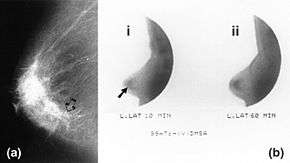 Mammography (left) and DMSA scintimammography (right) images of 4.5cm breast carcinoma | |
| Synonyms | Nuclear medicine breast imaging Breast specific gamma imaging Breast scintigraphy Molecular breast imaging |
| ICD-10-PCS | CH1 |
| ICD-9-CM | 92.19 |
| HCPCS-L2 | S8080 |
Scintimammography is not used for screening or in place of a mammogram. Rather, it is used when the detection of breast abnormalities is not possible or not reliable on the basis of mammography and ultrasound.[2]
Procedure
In the scintimammography procedure, a woman receives an injection of a small amount of a radioactive substance which is taken up by cancer cells, and a gamma camera is used to take pictures of the breasts.
Technetium-99m sestamibi (99mTc-MIBI) is the most commonly used radiopharmaceutical.[3][4] When performed with MIBI the procedure may also be known as a Miraluma test.[5] The typical dose is 740–1,110 megabecquerels (MBq) with the patient lying prone on the bed for imaging approximately 10 minutes after injection.[6]
99mTc-DMSA and 99mTc-MDP may also been used, but provide less sensitive results.[7]
Equipment
Breast-specific gamma cameras have been developed with a smaller field of view than conventional cameras, allowing higher resolution imagery and compression of the breast as in x-ray mammography (which improves detection of smaller lesions).[8][9]
References
- Liberman, Moishe; Sampalis, Fotini; Mulder, David S.; Sampalis, John S. (2003). "Breast Cancer Diagnosis by Scintimammography: A Meta-analysis and Review of the Literature". Breast Cancer Research and Treatment. 80 (1): 115–126. doi:10.1023/A:1024417331304. ISSN 0167-6806. PMID 12889605.
- Taillefer, Raymond (January 1999). "The role of 99mTc-sestamibi and other conventional radiopharmaceuticals in breast cancer diagnosis". Seminars in Nuclear Medicine. 29 (1): 16–40. doi:10.1016/S0001-2998(99)80027-0. PMID 9990681.
- Salvatore, Marco; Del Vecchio, Silvana (May 1998). "Dynamic imaging: scintimammography". European Journal of Radiology. 27: S259–S264. doi:10.1016/S0720-048X(98)00072-2.
- Nass, Sharyl J.; Henderson, I. Craig; Cancer, Institute of Medicine (U.S.). Committee on Technologies for the Early Detection of Breast (2001). Mammography and beyond: developing technologies for the early detection of breast cancer. National Academies Press. pp. 106–. ISBN 978-0-309-07283-0. Retrieved 17 July 2011.
- Tartar, Marie; Comstock, Christopher E.; Kipper, Michael S. (2008). Breast cancer imaging: a multidisciplinary, multimodality approach. Elsevier Health Sciences. pp. 88–. ISBN 978-0-323-04677-0. Retrieved 18 July 2011.
- "Procedure Guideline for Breast Scintigraphy" (PDF). Society of Nuclear Medicine. 2004. Cite journal requires
|journal=(help) - Piccart, Martine; Wood, William C.; Hung, Chie-Mien; Solin, Lawrence J.; Cardoso, Fatima (2007-03-07). Breast Cancer Management and Molecular Medicine. Springer Science & Business Media. p. 36. ISBN 9783540282655.
- Fass, Leonard (August 2008). "Imaging and cancer: A review". Molecular Oncology. 2 (2): 115–152. doi:10.1016/j.molonc.2008.04.001. PMC 5527766. PMID 19383333.
- Goldsmith, S. J.; Parsons, W.; Guiberteau, M. J.; Stern, L. H.; Lanzkowsky, L.; Weigert, J.; Heston, T. F.; Jones, E.; Buscombe, J.; Stabin, M. G. (5 November 2010). "SNM Practice Guideline for Breast Scintigraphy with Breast-Specific γ-Cameras 1.0". Journal of Nuclear Medicine Technology. 38 (4): 219–224. doi:10.2967/jnmt.110.082271. PMID 21057112.
External links
- Scintimammography entry in the public domain NCI Dictionary of Cancer Terms
![]()
| X-ray/ Radiography |
| ||||||||||||
|---|---|---|---|---|---|---|---|---|---|---|---|---|---|
| MRI | |||||||||||||
| Ultrasound | |||||||||||||
| Radionuclide |
| ||||||||||||
| Optical/Laser | |||||||||||||
| Thermography |
| ||||||||||||
| Target conditions |
| ||||||||||||
| |||||||||||||