Microvillous inclusion disease
Microvillus inclusion disease, also known as Davidson's disease, congenital microvillus atrophy and, less specifically, microvillus atrophy (note: microvillus is often misspelled as microvillous), is a rare genetic disorder of the small intestine that is inherited in an autosomal recessive pattern.[1][2]
| Microvillus inclusion disease | |
|---|---|
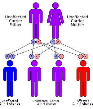 | |
| Microvillous inclusion disease has an autosomal recessive pattern of inheritance. |
Presentation
It is characterized by chronic, intractable diarrhea in new-born infants, starting in the first few days of life.[3] This results in metabolic acidosis and severe dehydration. Pregnancy and birth are usually normal.
Pathophysiology
It is caused by a congenital atrophy of apical microvilli and intracellular accumulation of apical enzymes in the epithelial cells of the small intestine.[4]
Diagnosis
Prenatal screening in utero is currently offered by several medical centers since the gene(s) involved in the disease were recently discovered to be MYO5B;[5][6] Diagnosis is typically made by biopsy of the small intestine.[1]
Biopsy
The appearance of microvillous inclusion disease on light microscopy is similar to celiac sprue; however, it usually lacks the intraepithelial lymphocytic infiltration characteristic of celiac sprue and stains positive for carcinoembryonic antigen (CEA).[2] The definitive diagnosis is dependent on electron microscopy.[7]
Differential diagnosis
The differential diagnosis of chronic and intractable diarrhea is:[8]
- Intestinal epithelial dysplasia
- Syndromatic diarrhea
- Immunoinflammatory enteropathy
Prognosis
It is nearly always fatal unless, like short bowel syndrome patients, treated with parenteral nutrition or an intestinal transplant.[3] The patient is often classified as being in "intestinal failure" and treated with the cohort of patients known as "short bowel syndrome" patients.
One patient from the UK was documented as achieving nutritional independence at age 3.[9]
On 26 June 2009 a six-year-old girl with microvillus inclusion disease became the third person in the UK to die of swine flu. This was attributed to her weakened immune system.[10]
Genetic prevalence
Microvillus inclusion disease is extremely rare, however, no prevalence data have been published. An estimate of a few hundred children with the disease in Europe has been made but no time frame to which this count applies is given. Countries with a higher degrees of consanguinity experience higher prevalence rates due to its autosomal recessive transmission.[11]
History
Microvillus inclusion disease was first described in 1978 by Davidson et al.[12] It was originally described as familial enteropathy.
References
- Chehade, Mirna; Sicherer, Scott H (2005). "Infantile food protein-induced enterocolitis syndrome". In David, Timothy J (ed.). Recent Advances in Paediatrics 22. London: Royal Society of Medicine Press. p. 140. ISBN 1-85315-572-1.
- Mills SE, Carter D, Greenson JK, Oberman HA, Reuter V, Stoler MH. Sternberg's Diagnostic Surgical Pathology. 4th Ed. Lippincott Williams & Wilkins. Copyright 2004. ISBN 978-0-7817-4051-7.
- Salvatore, S.; Hauser, B.; Vandenplas, Y. (2007). "Chronic enteropathy and feeding". In Cooke, Richard J.; Vandenplas, Yvan; Wahn, Ulrich (eds.). Nutrition Support for Infants and Children at Risk. Basel, Switzerland; New York: Karger. p. 123. ISBN 3-8055-8194-7.
- Arpin, M.; Crepaldi, T.; Louvard, D. (1999). "Cross-talk between Apical and Basolateral Domains of Epithelial Cells Regulates Microvillus Assembly". In Birchmeier, Walter; Birchmeier, Carmen (eds.). Epithelial Morphogenesis in Development and Disease. Amsterdam: Harwood Academic. p. 104. ISBN 90-5702-419-5.
- Mueller T; Hess, MW; Schiefermeier, N; Pfaller, K; Ebner, HL; Heinz-Erian, P; Ponstingl, H; Partsch, J; et al. (2008). "MYO5B mutations cause microvillus inclusion disease and disrupt epithelial cell polarity". Nat Genet. 40 (10): 1163–5. doi:10.1038/ng.225. PMID 18724368.
- Szperl A, Golachowska M, Rings E, IJzendoorn S, et al. (2011). "Functional characterization of mutations in the myosin Vb gene associated with microvillus inclusion disease". J Ped Gastroenterol Nutr. 52 (3): 307–13. doi:10.1097/MPG.0b013e3181eea177. PMC 3058815. PMID 21206382.
- Kennea N, Norbury R, Anderson G, Tekay A (2001). "Congenital microvillous inclusion disease presenting as antenatal bowel obstruction". Ultrasound Obstet Gynecol. 17 (2): 172–4. doi:10.1046/j.1469-0705.2001.00211.x. PMID 11251929.
- Ruemmele FM (2007). "Chronic enteropathy: molecular basis". Nestle Nutr Workshop Ser Pediatr Program. Series Set, 2007. 59: 73–85, discussion 85–8. doi:10.1159/000098514. ISBN 3-8055-8194-7. PMID 17245092.
- Croft NM; Howatson, AG; Ling, SC; Nairn, L; Evans, TJ; Weaver, LT (2000). "Microvillous inclusion disease: An evolving Condition". J Pediatr Gastroenterol Nutr. 31 (2): 185–189. doi:10.1097/00005176-200008000-00019. PMID 10941974.
- "Swine flu girl 'had tough life'". BBC News. 30 June 2009. Retrieved 12 May 2010.
- Ruemmele, Frank M; Schmitz, Jacques; Goulet, Olivier (2006-06-26). "Microvillous inclusion disease (microvillous atrophy)". Orphanet Journal of Rare Diseases. 1: 22. doi:10.1186/1750-1172-1-22. ISSN 1750-1172. PMC 1523325. PMID 16800870.
- Davidson GP, Cutz E, Hamilton JR, Gall DG (1978). "Familial enteropathy: a syndrome of protracted diarrhea from birth, failure to thrive, and hypoplastic villus atrophy". Gastroenterology. 75 (5): 783–90. doi:10.1016/0016-5085(78)90458-4. PMID 100367.
External links
| Classification | |
|---|---|
| External resources |