Microchimerism
Microchimerism is the presence of a small number of cells that originate from another individual and are therefore genetically distinct from the cells of the host individual. This phenomenon may be related to certain types of autoimmune diseases; however, the mechanisms responsible for this relationship are unclear.
Types
Human
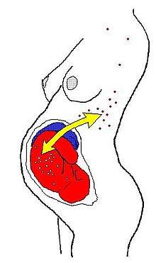
In humans (and perhaps in all placentals), the most common form is fetomaternal microchimerism (also known as fetal cell microchimerism or fetal chimerism) whereby cells from a fetus pass through the placenta and establish cell lineages within the mother. Fetal cells have been documented to persist and multiply in the mother for several decades.[1][2] The exact phenotype of these cells is unknown, although several different cell types have been identified, such as various immune lineages, mesenchymal stem cells, and placental-derived cells.[3] A 2012 study at the Fred Hutchinson Cancer Research Center, Seattle, has detected cells with the Y chromosome in multiple areas of the brains of deceased women.[4]
Fetomaternal microchimerism occurs during pregnancy and shortly after giving birth for most women. However, not all women who have had children contain fetal cells. Studies suggest that fetomaternal microchimerism could be influenced by killer-cell immunoglobin-like (KIR) ligands.[5] Lymphocytes also influence the development of persisting fetomaternal microchimerism since natural killer cells compose about 70% of lymphocytes in the first trimester of pregnancy. KIR patterns on maternal natural killer cells of the mother and KIR ligands on the fetal cells could have an effect on fetomaternal microchimerism. In one study, mothers with KIR2DS1 exhibited higher levels of fetomaternal microchimerism compared to mothers who were negative for this activating KIR.[5]
The potential health consequences of these cells are unknown. One hypothesis is that these fetal cells might trigger a graft-versus-host reaction leading to autoimmune disease. This offers a potential explanation for why many autoimmune diseases are more prevalent in middle-aged women.[6] Another hypothesis is that fetal cells home to injured or diseased maternal tissue where they act as stem cells and participate in repair.[7][8] It is also possible that the fetal cells are merely innocent bystanders and have no effect on maternal health.[9]
After giving birth, about 50–75% of women carry fetal immune cell lines. Maternal immune cells are also found in the offspring yielding in maternal→fetal microchimerism, though this phenomenon is about half as frequent as the former.[10]
Microchimerism had also been shown to exist after blood transfusions to a severely immunocompromised population of patients who suffered trauma.[11]
Other possible sources of microchimerism include gestation,[12] an individual's older sibling, twin sibling, or vanished twin, with the cells being received in utero. Fetal-maternal microchimerism is especially prevalent after abortion or miscarriage.[13] It is hypothesized that unprotected intercourse with ejaculation may be a potential source of microchimerism in rare cases.[14][15]
Animal
Microchimerism occurs in most pairs of twins in cattle. In cattle (and other bovines), the placentae of fraternal twins usually fuse and the twins share blood circulation, resulting in exchange of cell lines. If the twins are a male–female pair, then XX/XY microchimerism results, and male hormones partially masculinize the heifer (female), creating a martin heifer or freemartin. Freemartins appear female, but are infertile and so cannot be used for breeding or dairy production. Microchimerism provides a method of diagnosing the condition, because male genetic material can be detected in a blood sample.[16]
Fetomaternal microchimerism in the brain
Several studies have identified male DNA in both the human and mouse brains of mothers following pregnancy of a male fetus [17] [18] It has been suggested that the fetal-derived cells can differentiate into those capable of presenting neurotypical immunomarkers on their surface [19]. Fetal-derived cells in the brain appear to be able to persist in the human brain decades after a woman gives birth [20]. There has been no strong evidence to say microchimarism of the maternal brain leads to disease, however, Parkingson’s disease correlates with a higher incidence of brain microchimeras [Zeng]. Alzheimer’s disease studies support nearly the opposite correlation, the more fetal-derived cells present, the lower the chance of the patient having had Alzheimer’s . [21].
Relationship with autoimmune diseases and breast cancer
Microchimerism has been implicated in autoimmune diseases. Independent studies repeatedly suggested that microchimeric cells of fetal origin may be involved in the pathogenesis of systemic sclerosis.[2][22] Moreover, microchimeric cells of maternal origin may be involved in the pathogenesis of a group of autoimmune diseases found in children, i.e. juvenile idiopathic inflammatory myopathies (one example would be juvenile dermatomyositis).[23] Microchimerism has now been further implicated in other autoimmune diseases, including systemic lupus erythematosus.[24] Contrarily, an alternative hypothesis on the role of microchimeric cells in lesions is that they may be facilitating tissue repair of the damaged organ.[25]
Moreover, fetal immune cells have also been frequently found in breast cancer stroma as compared to samples taken from healthy women. It is not clear, however, whether fetal cell lines promote the development of tumors or, contrarily, protect women from developing breast carcinoma.[26][27]
Systemic lupus erythematosus
The presence of fetal cells in mothers can be associated with benefits when it comes to certain autoimmune diseases. In particular, male fetal cells are related to helping mothers with systemic lupus erythematosus. When kidney biopsies were taken from patients with lupus nephritis, DNA was extracted and run with PCR. The male fetal DNA was quantified and the presence of specific Y chromosome sequences were found. Women with lupus nephritis containing male fetal cells in their kidney biopsies exhibited better renal system functioning. Levels of serum creatinine, which is related to kidney failure, were low in mothers with high levels of male fetal cells.[28] In contrast, women without male fetal cells who had lupus nephritis showed a more serious form of glomerulonephritis and higher levels of serum creatinine.[28]
The specific role that fetal cells play in microchimerism related to certain autoimmune diseases is not fully understood. However, one hypothesis states that these cells supply antigens, causing inflammation and triggering the release of different foreign antigens.[28] This would trigger autoimmune disease instead of serving as a therapeutic. A different hypothesis states that fetal microchimeric cells are involved in repairing tissues. When tissues get inflamed, fetal microchimeric cells go to the damaged site and aid in repair and regeneration of the tissue.[28]
Thyroid disease
Fetal maternal microchimerism may be related to autoimmune thyroid diseases. There have been reports of fetal cells in the lining of the blood and thyroid glands of patients with autoimmune thyroid disease. These cells could become activated after delivery of the baby after immune suppression in the mother is lost, suggesting a role of fetal cells in the pathogenesis of such diseases.[29] Two types of thyroid disease, Hashimoto’s Thyroiditis (HT) and Graves’ Disease (GD) show similarities to graft vs host disease which occurs after hematopoietic stem cell transplants. Fetal cells colonize maternal tissues like the thyroid gland and are able to survive many years postpartum. These fetal microchimeric cells in the thyroid show up in the blood of women affected by thyroid diseases.[29]
Breast cancer
Pregnancy has a positive effect on the prognosis of breast cancer according to several studies [30][31][32] also it apparently increases the chance of survival after diagnosis of this tumor disease.[33] Possible positive effects of pregnancy could be explained by the persistence of fetal cells in the blood and maternal tissues.[34]
Fetal cells are probably actively migrating from peripheral blood into the tumor tissue [35] where they are preferentially settled in the tumor stroma [36] and their concentration decreases as they get closer to the healthy breast tissue.[37] There are two suggested mechanisms by which the fetal cells could have the positive effect on the breast cancer prognosis. First mechanism suggests that fetal cells only oversee cancer cells and they attract components of the immune system if needed. The second option is that the down-regulation of the immune system induced by the presence of fetal cells could ultimately lead to cancer prevention, because women in whom FMC is present produce lower concentrations of inflammatory mediators, which may lead to the development of neoplastic tissue.[38]
The effect also depends on the level of microchimerism: Hyperchimerism (too high a degree of microchimerism) can, as well as hypochimerism (low rate of microchimerism), be related to the negative effect of FMC and thus can promote worse prognosis of breast cancer.[39][40] Apparently, women with breast cancer may fail in the process of obtaining and maintaining allogeneic fetal cells. Low concentration and / or complete absence of fetal cells could indicate a predisposition to development of the malignant process.
Stem cells
Animal models
Fetomaternal microchimerism has been shown in experimental investigations of whether fetal cells can cross the blood brain barrier in mice. The properties of these cells allow them to cross the blood brain barrier and target injured brain tissue.[41] This mechanism is possible because umbilical cord blood cells express some proteins similar to neurons. When these umbilical cord blood cells are injected in rats with brain injury or stroke, they enter the brain and express certain nerve cell markers. Due to this process, fetal cells could enter the brain during pregnancy and become differentiated into neural cells. Fetal microchimerism can occur in the maternal mouse brain, responding to certain cues in the maternal body.[41]
Health implications
Fetal microchimerism could have an implication on maternal health. Isolating cells in cultures can alter the properties of the stem cells, but in pregnancy the effects of fetal stem cells can be investigated without in vitro cultures. Once characterized and isolated, fetal cells that are able to cross the blood brain barrier could impact certain procedures.[41] For example, isolating stem cells can be accomplished through taking them from sources like the umbilical cord. These fetal stem cells can be used in intravenous infusion to repair the brain tissue. Hormonal changes in pregnancy alter neurogenesis, which could create favorable environments for fetal cells to respond to injury.[41]
The true function on fetal cells in mothers is not fully known, however, there have been reports of positive and negative health effects. The sharing of genes between the fetus and mother may lead to benefits. Due to not all genes being shared, health complications may arise as a result of resource allocation.[42] During pregnancy, fetal cells are able to manipulate the maternal system to draw resources from the placenta, while the maternal system tries to limit it.[42]
See also
- Chimerism
- Allotransplantation
- Telegony
- Epigenetics
- Cell-free fetal DNA
References
- Bianchi DW, Zickwolf GK, Weil GJ, Sylvester S, DeMaria MA (January 1996). "Male fetal progenitor cells persist in maternal blood for as long as 27 years postpartum". Proceedings of the National Academy of Sciences of the United States of America. 93 (2): 705–8. Bibcode:1996PNAS...93..705B. doi:10.1073/pnas.93.2.705. PMC 40117. PMID 8570620.
- Evans PC, Lambert N, Maloney S, Furst DE, Moore JM, Nelson JL (March 1999). "Long-term fetal microchimerism in peripheral blood mononuclear cell subsets in healthy women and women with scleroderma". Blood. 93 (6): 2033–7. doi:10.1182/blood.V93.6.2033.406k18_2033_2037. PMID 10068676.
- Pritchard S, Wick HC, Slonim DK, Johnson KL, Bianchi DW (August 2012). "Comprehensive analysis of genes expressed by rare microchimeric fetal cells in the maternal mouse lung". Biology of Reproduction. 87 (2): 42. doi:10.1095/biolreprod.112.101147. PMC 3431427. PMID 22674387.
- Chan WF, Gurnot C, Montine TJ, Sonnen JA, Guthrie KA, Nelson JL (26 September 2012). "Male microchimerism in the human female brain". PLOS ONE. 7 (9): e45592. Bibcode:2012PLoSO...745592C. doi:10.1371/journal.pone.0045592. PMC 3458919. PMID 23049819.
- Kruchen A, Stahl T, Gieseke F, Binder TM, Oezcan Z, Meisel R, et al. (2014). "Fetomaternal Microchimerism Is Associated with Better Outcome in Haploidentical Hematopoietic Stem Cell Transplantation". Blood. 124 (21). Retrieved 3 December 2016.
- Nelson JL (February 1996). "Maternal-fetal immunology and autoimmune disease: is some autoimmune disease auto-alloimmune or allo-autoimmune?". Arthritis and Rheumatism. 39 (2): 191–4. doi:10.1002/art.1780390203. PMID 8849367.
- Khosrotehrani K, Johnson KL, Cha DH, Salomon RN, Bianchi DW (July 2004). "Transfer of fetal cells with multilineage potential to maternal tissue". JAMA. 292 (1): 75–80. doi:10.1001/jama.292.1.75. PMID 15238593.
- Nguyen Huu S, Oster M, Avril MF, Boitier F, Mortier L, Richard MA, Kerob D, Maubec E, Souteyrand P, Moguelet P, Khosrotehrani K, Aractingi S (February 2009). "Fetal microchimeric cells participate in tumour angiogenesis in melanomas occurring during pregnancy". The American Journal of Pathology. 174 (2): 630–7. doi:10.2353/ajpath.2009.080566. PMC 2630570. PMID 19147820.
- Johnson KL, Bianchi DW (2004). "Fetal cells in maternal tissue following pregnancy: what are the consequences?". Human Reproduction Update. 10 (6): 497–502. doi:10.1093/humupd/dmh040. PMID 15319378.
- Loubière LS, Lambert NC, Flinn LJ, Erickson TD, Yan Z, Guthrie KA, Vickers KT, Nelson JL (November 2006). "Maternal microchimerism in healthy adults in lymphocytes, monocyte/macrophages and NK cells". Laboratory Investigation; A Journal of Technical Methods and Pathology. 86 (11): 1185–92. doi:10.1038/labinvest.3700471. PMID 16969370.
- Reed W, Lee TH, Norris PJ, Utter GH, Busch MP (January 2007). "Transfusion-associated microchimerism: a new complication of blood transfusions in severely injured patients". Seminars in Hematology. 44 (1): 24–31. doi:10.1053/j.seminhematol.2006.09.012. PMID 17198844.
- Shree, Raj, et al. "Fetal Microchimerism by Mode of Delivery in Healthy Term Gestations." REPRODUCTIVE SCIENCES. Vol. 24. 2455 TELLER RD, THOUSAND OAKS, CA 91320 USA: SAGE PUBLICATIONS INC, 2017.
- Khosrotehrani K, Johnson KL, Lau J, Dupuy A, Cha DH, Bianchi DW (November 2003). "The influence of fetal loss on the presence of fetal cell microchimerism: a systematic review". Arthritis and Rheumatism. 48 (11): 3237–41. doi:10.1002/art.11324. PMID 14613289.
- Yan Z, Lambert NC, Guthrie KA, Porter AJ, Loubiere LS, Madeleine MM, Stevens AM, Hermes HM, Nelson JL (August 2005). "Male microchimerism in women without sons: quantitative assessment and correlation with pregnancy history". The American Journal of Medicine. 118 (8): 899–906. doi:10.1016/j.amjmed.2005.03.037. PMID 16084184.
- Müller AC, Jakobsen MA, Barington T, Vaag AA, Grunnet LG, Olsen SF, Kamper-Jørgensen M (October 2015). "Microchimerism of male origin in a cohort of Danish girls". Chimerism. 6 (4): 65–71. doi:10.1080/19381956.2016.1218583. PMC 5293315. PMID 27623703.
- A. Fujishiro, K. Kawakura, Y-I. Miyake, Y. Kaneda, "A fast, convenient diagnosis of the bovine freemartin syndrome using polymerase chain reaction", Theriogenology, 43 (5), pp 883–891 (1 April 1995)
- Zeng XX, Tan KH, Yeo A, Sasajala P, et al. 2010. Pregnancyassociated progenitor cells differentiate and mature into neurons in the maternal brain. Stem Cells Dev.
- Chan WFN, Gurnot C, Montine TJ, Sonnen JA, et al. 2012. Male microchimerism in the human female brain. PLoS One
- Zeng XX, Tan KH, Yeo A, Sasajala P, et al. 2010. Pregnancyassociated progenitor cells differentiate and mature into neurons in the maternal brain. Stem Cells Dev.
- Chan WFN, Gurnot C, Montine TJ, Sonnen JA, et al. 2012. Male microchimerism in the human female brain. PLoS One
- Chan WFN, Gurnot C, Montine TJ, Sonnen JA, et al. 2012. Male microchimerism in the human female brain. PLoS One
- Artlett CM, Smith JB, Jimenez SA (April 1998). "Identification of fetal DNA and cells in skin lesions from women with systemic sclerosis". The New England Journal of Medicine. 338 (17): 1186–91. doi:10.1056/NEJM199804233381704. PMID 9554859.
- Artlett CM, Ramos R, Jiminez SA, Patterson K, Miller FW, Rider LG (2000). "Chimeric cells of maternal origin in juvenile idiopathic inflammatory myopathies. Childhood Myositis Heterogeneity Collaborative Group". Lancet. 356 (9248): 2155–6. doi:10.1016/S0140-6736(00)03499-1. PMID 11191545.
- Johnson KL, McAlindon TE, Mulcahy E, Bianchi DW (September 2001). "Microchimerism in a female patient with systemic lupus erythematosus". Arthritis and Rheumatism. 44 (9): 2107–11. doi:10.1002/1529-0131(200109)44:9<2107::AID-ART361>3.0.CO;2-9. PMID 11592373.
- Gilliam AC (February 2006). "Microchimerism and skin disease: true-true unrelated?". The Journal of Investigative Dermatology. 126 (2): 239–41. doi:10.1038/sj.jid.5700061. PMID 16418731.
- Gadi VK, Nelson JL (October 2007). "Fetal microchimerism in women with breast cancer". Cancer Research. 67 (19): 9035–8. doi:10.1158/0008-5472.CAN-06-4209. PMID 17909006.
- Dubernard G, Aractingi S, Oster M, Rouzier R, Mathieu MC, Uzan S, Khosrotehrani K (2008). "Breast cancer stroma frequently recruits fetal derived cells during pregnancy". Breast Cancer Research. 10 (1): R14. doi:10.1186/bcr1860. PMC 2374970. PMID 18271969.
- Florim GM, Caldas HC, de Melo JC, Baptista MA, Fernandes IM, Savoldi-Barbosa M, Goldman GH, Abbud-Filho M (April 2015). "Fetal microchimerism in kidney biopsies of lupus nephritis patients may be associated with a beneficial effect". Arthritis Research & Therapy. 17: 101. doi:10.1186/s13075-015-0615-4. PMC 4416327. PMID 25889410.
- Lepez T, Vandewoestyne M, Deforce D (20 May 2013). "Fetal microchimeric cells in autoimmune thyroid diseases: harmful, beneficial or innocent for the thyroid gland?". Chimerism. 4 (4): 111–8. doi:10.4161/chim.25055. PMC 3921191. PMID 23723083.
- Rosenberg L, Thalib L, Adami HO, Hall P (September 2004). "Childbirth and breast cancer prognosis". International Journal of Cancer. 111 (5): 772–6. doi:10.1002/ijc.20323. PMID 15252849.
- Olson, Sara H.; Zauber, Ann G.; Tang, Jian; Harlap, Susan (November 1998). "Relation of Time since Last Birth and Parity to Survival of Young Women with Breast Cancer". Epidemiology. 9 (6): 669–671. doi:10.1097/00001648-199811000-00019. ISSN 1044-3983.
- Anderson, Penny R.; Hanlon, Alexandra L.; Freedman, Gary M.; Nicolaou, Nicos (August 2004). "Parity Confers Better Prognosis in Older Women with Early-Stage Breast Cancer Treated with Breast-Conserving Therapy". Clinical Breast Cancer. 5 (3): 225–231. doi:10.3816/cbc.2004.n.026. ISSN 1526-8209. PMID 15335456.
- Warren Andersen S, Newcomb PA, Hampton JM, Titus-Ernstoff L, Egan KM, Trentham-Dietz A (December 2011). "Reproductive factors and histologic subtype in relation to mortality after a breast cancer diagnosis". Breast Cancer Research and Treatment. 130 (3): 975–80. doi:10.1007/s10549-011-1666-0. PMC 4306414. PMID 21769659.
- Bianchi, D. W.; Zickwolf, G. K.; Weil, G. J.; Sylvester, S.; DeMaria, M. A. (1996-01-23). "Male fetal progenitor cells persist in maternal blood for as long as 27 years postpartum". Proceedings of the National Academy of Sciences. 93 (2): 705–708. Bibcode:1996PNAS...93..705B. doi:10.1073/pnas.93.2.705. ISSN 0027-8424. PMC 40117. PMID 8570620.
- Dubernard G, Oster M, Chareyre F, Antoine M, Rouzier R, Uzan S, Aractingi S, Khosrotehrani K (March 2009). "Increased fetal cell microchimerism in high grade breast carcinomas occurring during pregnancy". International Journal of Cancer. 124 (5): 1054–9. doi:10.1002/ijc.24036. PMID 19065666.
- Dubernard G, Aractingi S, Oster M, Rouzier R, Mathieu MC, Uzan S, Khosrotehrani K (February 2008). "Breast cancer stroma frequently recruits fetal derived cells during pregnancy". Breast Cancer Research. 10 (1): R14. doi:10.1186/bcr1860. PMC 2374970. PMID 18271969.
- Nemescu D, Ursu RG, Nemescu ER, Negura L (2016-01-25). "Heterogeneous Distribution of Fetal Microchimerism in Local Breast Cancer Environment". PLOS ONE. 11 (1): e0147675. Bibcode:2016PLoSO..1147675N. doi:10.1371/journal.pone.0147675. PMC 4726590. PMID 26808509.
- Coussens LM, Werb Z (2002-12-19). "Inflammation and cancer". Nature. 420 (6917): 860–7. Bibcode:2002Natur.420..860C. doi:10.1038/nature01322. PMC 2803035. PMID 12490959.
- Gadi VK, Malone KE, Guthrie KA, Porter PL, Nelson JL (March 2008). "Case-control study of fetal microchimerism and breast cancer". PLOS ONE. 3 (3): e1706. Bibcode:2008PLoSO...3.1706G. doi:10.1371/journal.pone.0001706. PMC 2248618. PMID 18320027.
- Dhimolea E, Denes V, Lakk M, Al-Bazzaz S, Aziz-Zaman S, Pilichowska M, Geck P (August 2013). "High male chimerism in the female breast shows quantitative links with cancer". International Journal of Cancer. 133 (4): 835–42. doi:10.1002/ijc.28077. PMID 23390035.
- Tan XW, Liao H, Sun L, Okabe M, Xiao ZC, Dawe GS (1 November 2005). "Fetal microchimerism in the maternal mouse brain: a novel population of fetal progenitor or stem cells able to cross the blood-brain barrier?". Stem Cells. 23 (10): 1443–52. doi:10.1634/stemcells.2004-0169. PMID 16091558.
- Boddy AM, Fortunato A, Wilson Sayres M, Aktipis A (October 2015). "Fetal microchimerism and maternal health: a review and evolutionary analysis of cooperation and conflict beyond the womb". BioEssays. 37 (10): 1106–18. doi:10.1002/bies.201500059. PMC 4712643. PMID 26316378.
Further reading
- Kinder JM, Stelzer IA, Arck PC, Way SS (August 2017). "Immunological implications of pregnancy-induced microchimerism". Nature Reviews. Immunology. 17 (8): 483–494. doi:10.1038/nri.2017.38. PMC 5532073. PMID 28480895.
- Müller AC, Jakobsen MA, Barington T, Vaag AA, Grunnet LG, Olsen SF, Kamper-Jørgensen M (October 2015). "Microchimerism of male origin in a cohort of Danish girls". Chimerism. 6 (4): 65–71. doi:10.1080/19381956.2016.1218583. PMC 5293315. PMID 27623703.
- Gammill HS, Nelson JL (2010). "Naturally acquired microchimerism". The International Journal of Developmental Biology. 54 (2–3): 531–43. doi:10.1387/ijdb.082767hg. PMC 2887685. PMID 19924635.