Medical physics
Medical physics (also called biomedical physics, medical biophysics, applied physics in medicine, physics applications in medical science, radiological physics or hospital radio-physics) is, in general, the application of physics concepts, theories, and methods to medicine or healthcare. Medical physics departments may be found in hospitals or universities.
In the case of hospital work, the term medical physicist is the title of a specific healthcare profession, usually working within a hospital. Medical physicists are often found in the following healthcare specialties: diagnostic and interventional radiology (also known as medical imaging), nuclear medicine, radiation protection and radiation oncology.
University departments are of two types. The first type are mainly concerned with preparing students for a career as a hospital medical physicist and research focuses on improving the practice of the profession. A second type (increasingly called 'biomedical physics') has a much wider scope and may include research in any applications of physics to medicine from the study of biomolecular structure to microscopy and nanomedicine. For example, physicist Richard Feynman theorized about the future of nanomedicine. He wrote about the idea of a medical use for biological machines (see nanobiotechnology). Feynman and Albert Hibbs suggested that certain repair machines might one day be reduced in size to the point that it would be possible to (as Feynman put it) "swallow the doctor". The idea was discussed in Feynman's 1959 essay There's Plenty of Room at the Bottom.[1]
Mission statement of medical physicists
In the case of hospital medical physics departments, the mission statement for medical physicists as adopted by the European Federation of Medical Physicists is the following:[2][3]
"Medical Physicists will contribute to maintaining and improving the quality, safety and cost-effectiveness of healthcare services through patient-oriented activities requiring expert action, involvement or advice regarding the specification, selection, acceptance testing, commissioning, quality assurance/control and optimised clinical use of medical devices and regarding patient risks and protection from associated physical agents (e.g., x-rays, electromagnetic fields, laser light, radionuclides) including the prevention of unintended or accidental exposures; all activities will be based on current best evidence or own scientific research when the available evidence is not sufficient. The scope includes risks to volunteers in biomedical research, carers and comforters. The scope often includes risks to workers and public particularly when these impact patient risk"
The term "physical agents" refers to ionising and non-ionising electromagnetic radiations, static electric and magnetic fields, ultrasound, laser light and any other Physical Agent associated with medical e.g., x-rays in computerised tomography (CT), gamma rays/radionuclides in nuclear medicine, magnetic fields and radio-frequencies in magnetic resonance imaging (MRI), ultrasound in ultrasound imaging and Doppler measurements.
This mission includes the following 11 key activities:
- Scientific problem solving service: Comprehensive problem solving service involving recognition of less than optimal performance or optimised use of medical devices, identification and elimination of possible causes or misuse, and confirmation that proposed solutions have restored device performance and use to acceptable status. All activities are to be based on current best scientific evidence or own research when the available evidence is not sufficient.
- Dosimetry measurements: Measurement of doses suffered by patients, volunteers in biomedical research, carers, comforters and persons subjected to non-medical imaging exposures (e.g., for legal or employment purposes); selection, calibration and maintenance of dosimetry related instrumentation; independent checking of dose related quantities provided by dose reporting devices (including software devices); measurement of dose related quantities required as inputs to dose reporting or estimating devices (including software). Measurements to be based on current recommended techniques and protocols. Includes dosimetry of all physical agents.
- Patient safety/risk management (including volunteers in biomedical research, carers, comforters and persons subjected to non-medical imaging exposures. Surveillance of medical devices and evaluation of clinical protocols to ensure the ongoing protection of patients, volunteers in biomedical research, carers, comforters and persons subjected to non-medical imaging exposures from the deleterious effects of physical agents in accordance with the latest published evidence or own research when the available evidence is not sufficient. Includes the development of risk assessment protocols.
- Occupational and public safety/risk management (when there is an impact on medical exposure or own safety). Surveillance of medical devices and evaluation of clinical protocols with respect to protection of workers and public when impacting the exposure of patients, volunteers in biomedical research, carers, comforters and persons subjected to non-medical imaging exposures or responsibility with respect to own safety. Includes the development of risk assessment protocols in conjunction with other experts involved in occupational / public risks.
- Clinical medical device management: Specification, selection, acceptance testing, commissioning and quality assurance/ control of medical devices in accordance with the latest published European or International recommendations and the management and supervision of associated programmes. Testing to be based on current recommended techniques and protocols.
- Clinical involvement: Carrying out, participating in and supervising everyday radiation protection and quality control procedures to ensure ongoing effective and optimised use of medical radiological devices and including patient specific optimization.
- Development of service quality and cost-effectiveness: Leading the introduction of new medical radiological devices into clinical service, the introduction of new medical physics services and participating in the introduction/development of clinical protocols/techniques whilst giving due attention to economic issues.
- Expert consultancy: Provision of expert advice to outside clients (e.g., clinics with no in-house medical physics expertise).
- Education of healthcare professionals (including medical physics trainees: Contributing to quality healthcare professional education through knowledge transfer activities concerning the technical-scientific knowledge, skills and competences supporting the clinically effective, safe, evidence-based and economical use of medical radiological devices. Participation in the education of medical physics students and organisation of medical physics residency programmes.
- Health technology assessment (HTA): Taking responsibility for the physics component of health technology assessments related to medical radiological devices and /or the medical uses of radioactive substances/sources.
- Innovation: Developing new or modifying existing devices (including software) and protocols for the solution of hitherto unresolved clinical problems.
Medical biophysics and biomedical physics
Some education institutions house departments or programs bearing the title "medical biophysics" or "biomedical physics" or "applied physics in medicine". Generally, these fall into one of two categories: interdisciplinary departments that house biophysics, radiobiology, and medical physics under a single umbrella;[4][5][6] and undergraduate programs that prepare students for further study in medical physics, biophysics, or medicine.[7][8]
Areas of specialty
The International Organization for Medical Physics (IOMP) recognizes main areas of medical physics employment and focus.[9][10] These are:
Medical imaging physics
.gif)
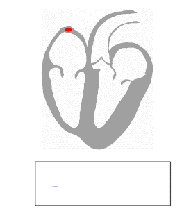
Medical imaging physics is also known as diagnostic and interventional radiology physics. Clinical (both "in-house" and "consulting") physicists[11] typically deal with areas of testing, optimization, and quality assurance of diagnostic radiology physics areas such as radiographic X-rays, fluoroscopy, mammography, angiography, and computed tomography, as well as non-ionizing radiation modalities such as ultrasound, and MRI. They may also be engaged with radiation protection issues such as dosimetry (for staff and patients). In addition, many imaging physicists are often also involved with nuclear medicine systems, including single photon emission computed tomography (SPECT) and positron emission tomography (PET). Sometimes, imaging physicists may be engaged in clinical areas, but for research and teaching purposes,[12] such as quantifying intravascular ultrasound as a possible method of imaging a particular vascular object.
Radiation therapeutic physics
Radiation therapeutic physics is also known as radiotherapy physics or radiation oncology physics. The majority of medical physicists currently working in the US, Canada, and some western countries are of this group. A radiation therapy physicist typically deals with linear accelerator (Linac) systems and kilovoltage x-ray treatment units on a daily basis, as well as other modalities such as TomoTherapy, gamma knife, cyberknife, proton therapy, and brachytherapy.[13][14][15] The academic and research side of therapeutic physics may encompass fields such as boron neutron capture therapy, sealed source radiotherapy, terahertz radiation, high-intensity focused ultrasound (including lithotripsy), optical radiation lasers, ultraviolet etc. including photodynamic therapy, as well as nuclear medicine including unsealed source radiotherapy, and photomedicine, which is the use of light to treat and diagnose disease.
Nuclear medicine physics
Nuclear medicine is a branch of medicine that uses radiation to provide information about the functioning of a person's specific organs or to treat disease. The thyroid, bones, heart, liver and many other organs can be easily imaged, and disorders in their function revealed. In some cases radiation sources can be used to treat diseased organs, or tumours. Five Nobel laureates have been intimately involved with the use of radioactive tracers in medicine. Over 10,000 hospitals worldwide use radioisotopes in medicine, and about 90% of the procedures are for diagnosis. The most common radioisotope used in diagnosis is technetium-99m, with some 30 million procedures per year, accounting for 80% of all nuclear medicine procedures worldwide.[16]
Health physics
Health physics is also known as radiation safety or radiation protection. Health physics is the applied physics of radiation protection for health and health care purposes. It is the science concerned with the recognition, evaluation, and control of health hazards to permit the safe use and application of ionizing radiation. Health physics professionals promote excellence in the science and practice of radiation protection and safety.
- Background radiation
- Radiation protection
- Dosimetry
- Health physics
- Radiological protection of patients
Non-ionizing Medical Radiation Physics
Some aspects of non-ionising radiation physics may be considered under radiation protection or diagnostic imaging physics. Imaging modalities include MRI, optical imaging and ultrasound. Safety considerations include these areas and lasers
Physiological measurement
Physiological measurements have also been used to monitor and measure various physiological parameters. Many physiological measurement techniques are non-invasive and can be used in conjunction with, or as an alternative to, other invasive methods. Measurement methods include electrocardiography Many of these areas may be covered by other specialities, for example medical engineering or vascular science.[17]
Healthcare informatics and computational physics
Other closely related fields to medical physics include fields which deal with medical data, information technology and computer science for medicine.
- Information and communication in medicine
- Medical informatics
- Image processing, display and visualization
- Computer-aided diagnosis
- Picture archiving and communication systems (PACS)
- Standards: DICOM, ISO, IHE
- Hospital information systems
- e-Health
- Telemedicine
- Digital operating room
- Workflow, patient-specific modeling
- Medicine on the Internet of Things
- Distant monitoring and telehomecare
Areas of research and academic development
Non-clinical physicists may or may not focus on the above areas from an academic and research point of view, but their scope of specialization may also encompass lasers and ultraviolet systems (such as photodynamic therapy), fMRI and other methods for functional imaging as well as molecular imaging, electrical impedance tomography, diffuse optical imaging, optical coherence tomography, and dual energy X-ray absorptiometry.
Legislative and advisory bodies
- ICRU: International Commission on Radiation Units and Measurements
- ICRP: International Commission on Radiological Protection
- NCRP: National Council on Radiation Protection & Measurements
- NRC: Nuclear Regulatory Commission
- FDA: Food and Drug Administration
- IAEA: International Atomic Energy Agency
- AMPI: Association of Medical Physicists of India
- AAPM: American Association of Physicists in Medicine
References
- Richard P. Feynman (December 1959). "There's Plenty of Room at the Bottom". Archived from the original on 2010-02-16. Retrieved 1 March 2010.
- Guibelalde E., Christofides S., Caruana C. J., Evans S. van der Putten W. (2012). Guidelines on the Medical Physics Expert' a project funded by the European Commission
- Caruana C.J., Christofides S., Hartmann G.H. (2014) European Federation of Organisations for Medical Physics (EFOMP) Policy Statement 12.1: Recommendations on Medical Physics Education and Training in Europe 2014 Physica Medica - European Journal of Medical Physics, 30:6, p598-603
- "Department of Medical Biophysics". utoronto.ca.
- "Medical Biophysics - Western University". uwo.ca. Archived from the original on 2013-07-03.
- UCLA Biomedical Physics Graduate Program
- "Welcome". wayne.edu. Archived from the original on 2013-08-12. Retrieved 2013-07-01.
- "Medical Physics". fresnostate.edu.
- "Medical Physics". International Organization for Medical Physics. Retrieved 21 October 2017.
- "AAPM Position Statements, Policies and Procedures - Details". aapm.org.
- "AAPM - What do Medical Physicists Do?". aapm.org.
- "Archived copy". Archived from the original on 2013-11-13. Retrieved 2013-11-13.CS1 maint: archived copy as title (link)
- Hill R, Healy B, Holloway L, Kuncic Z, Thwaites D, Baldock C (2014). "Advances in kilovoltage x-ray beam dosimetry". Physics in Medicine and Biology. 59 (6): R183–231. Bibcode:2014PMB....59R.183H. doi:10.1088/0031-9155/59/6/R183. PMID 24584183.CS1 maint: uses authors parameter (link)
- Thwaites DI, Tuohy JB (2006). "Back to the future: the history and development of the clinical linear accelerator". Physics in Medicine and Biology. 51 (13): R343–62. Bibcode:2006PMB....51R.343T. doi:10.1088/0031-9155/51/13/R20. PMID 16790912.CS1 maint: uses authors parameter (link)
- Mackie, T R (2006). "The history of tomotherapy". Physics in Medicine and Biology. 51 (13): R427–53. Bibcode:2006PMB....51R.427M. doi:10.1088/0031-9155/51/13/R24. PMID 16790916.
- "Radioisotopes in Medicine". World Nuclear Association. October 2017. Retrieved 21 October 2017.
- "Vascular science". NHS Health Careers. 25 March 2015. Retrieved 21 October 2017.
External links
| Wikimedia Commons has media related to Medical physics. |
- Human Health Campus, The official website of the International Atomic Energy Agency dedicated to Professionals in Radiation Medicine. This site is managed by the Division of Human Health, Department of Nuclear Sciences and Applications
- Australasian College of Physical Scientists and Engineers in Medicine (ACPSEM)
- Canadian Organization of Medical Physicist - Organisation canadienne des physiciens médicaux
- The American Association of Physicists in Medicine
- Romanian College of Medical Physicists
- medicalphysicsweb.org from the Institute of Physics
- AIP Medical Physics portal
- Institute of Physics & Engineering in Medicine (IPEM) - UK
- European Federation of Organizations for Medical Physics (EFOMP)
- International Organization for Medical Physics (IOMP)
| Authority control |
|
|---|