Medial palpebral ligament
The medial palpebral ligament (medial canthal tendon) is about 4 mm in length and 2 mm in breadth. Its anterior attachment is to the frontal process of the maxilla in front of the lacrimal groove, and its posterior attachment is the lacrimal bone. Laterally, it is attached to the tarsus of the upper and lower eyelids.
| Medial palpebral ligament | |
|---|---|
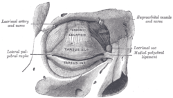 The tarsi and their ligaments. Right eye; front view. | |
| Details | |
| Identifiers | |
| Latin | Ligamentum palpebrale mediale, tendo oculi |
| TA | A15.2.07.041 |
| FMA | 323068 |
| Anatomical terminology | |
Crossing the lacrimal sac, it divides into two parts, upper and lower, each attached to the medial end of the corresponding tarsus.
As the ligament crosses the lacrimal sac, a strong aponeurotic lamina is given off from its posterior surface; this expands over the sac, and is attached to the posterior lacrimal crest.
See also
References
This article incorporates text in the public domain from page 381 of the 20th edition of Gray's Anatomy (1918)
| Authority control |
|---|
This article is issued from
Wikipedia.
The text is licensed under Creative
Commons - Attribution - Sharealike.
Additional terms may apply for the media files.