Deltoid ligament
The deltoid ligament (or medial ligament of talocrural joint) is a strong, flat, triangular band, attached, above, to the apex and anterior and posterior borders of the medial malleolus. The deltoid ligament is composed of: 1. Anterior tibiotalar ligament 2. Tibiocalcaneal ligament 3. Posterior tibiotalar ligament 4. Tibionavicular ligament. It consists of two sets of fibers, superficial and deep.
| Deltoid ligament | |
|---|---|
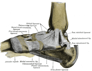 Ligaments of the medial aspect of the foot. | |
| Details | |
| From | Talus bone (tarsal bones) |
| To | Medial malleolus of the tibia |
| Identifiers | |
| Latin | Ligamentum collaterale mediale articulationis talocruralis, ligamentum deltoideum |
| TA | A03.6.10.003 |
| FMA | 44055 |
| Anatomical terminology | |
Superficial fibres
Of the superficial fibres,
- tibionavicular pass forward to be inserted into the tuberosity of the navicular bone, and immediately behind this they blend with the medial margin of the plantar calcaneonavicular ligament;
- tibiocalcaneal descend almost perpendicularly to be inserted into the whole length of the sustentaculum tali of the calcaneus;
- posterior tibiotalar from the posterior colliculus of the medial malleolus to the posteromedial of the talus
Deep fibres
The deep fibres (anterior tibiotalar) are attached from the anterior colliculus of the medial malleolus to the medial talus and medial tubercle
Coverings
The deltoid ligament is covered by the tendons of the tibialis posterior and flexor digitorum longus.
Additional Images
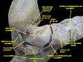 Ankle joint.Deep section.
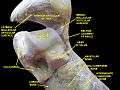 Ankle joint. Deep dissection.
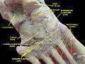 Ankle joint. Deep dissection.
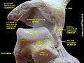 Ankle joint. Deep dissection.
References
This article incorporates text in the public domain from page 350 of the 20th edition of Gray's Anatomy (1918)
External links
- Deltoid_ligament at the Duke University Health System's Orthopedics program
- lljoints at The Anatomy Lesson by Wesley Norman (Georgetown University) (medialanklejoint)
- http://www.ithaca.edu/faculty/lahr/LE2000/ankle%20pics/5medankle-new.jpg
| Authority control |
|---|
- Thompson, Jon C. Netter's concise atlas of orthopaedic anatomy. Icon Learning Systems, 2002. 349-351