Magnetic resonance elastography
Magnetic resonance elastography (MRE) is a non-invasive medical imaging technique that measures the stiffness of soft tissues by generating shear waves in tissue, imaging their propagation using MRI, and processing the images to generate a stiffness map (elastogram).[1] It is one of the most commonly used elastography techniques.[2]
| Magnetic resonance elastography | |
|---|---|
| Medical diagnostics | |
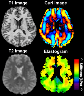 Magnetic resonance elastography of the brain. A T1 weighted anatomical image is shown in the top-left, and the corresponding T2 weighted image from the MRE data is shown in the bottom-left. The wave image used to make the elastogram is shown in the top-right, and the resulting elastogram is in the bottom-right. | |
| Purpose | measures the mechanical properties of soft tissues |
MRE was first described by Muthupillai et al. in 1995.[3] Because diseased tissues are often stiffer than the surrounding normal tissue, MRE has been applied to visualize a variety of disease processes which affect tissue stiffness in the liver, breast, brain, heart, and skeletal muscle.[1][4] For example, breast tumors are much harder than healthy fibroglandular tissue.[5] MRE is similar to palpation; however, whereas palpation is a qualitative technique performed by physicians, MRE is a quantitative technique performed with a radiologist.[1]
Applications
Liver
Liver fibrosis is a common result of many chronic liver diseases; progressive fibrosis can lead to cirrhosis. MRE of the liver provides quantitative maps of tissue stiffness over large regions of the liver. This non-invasive technique is able to detect increased stiffness of the liver parenchyma, which is a direct consequence of liver fibrosis. It helps to stage liver fibrosis or diagnose mild fibrosis with reasonable accuracy.[6][7][8][9]
Brain
MRE of the brain was first presented in the early 2000s.[10][11] Elastogram measures have been correlated with memory tasks,[12] fitness measures,[13] and progression of various neurodegenerative conditions. For example, regional and global decreases in brain viscoelasticity have been observed in Alzheimer’s disease[14][15] and multiple sclerosis.[16][17] It has been found that as the brain ages, it loses its viscoelastic integrity due to degeneration of neurons and oligodendrocytes.[18][19]
MRE may also have applications for understanding the adolescent brain. Recently, it was found that adolescents have regional differences in brain viscoelasticity relative to adults.[20][21]
MRE has also been applied to functional neuroimaging. Whereas functional magnetic resonance imaging (fMRI) infers brain activity by detecting relatively slow changes in blood flow, functional MRE is capable of detecting neuromechanical changes in the brain related to neuronal activity occurring on the 100-millisecond scale.[22]
References
- Mariappan YK, Glaser KJ, Ehman RL (2010). "Magnetic resonance elastography: a review". Clin Anat. 23 (5): 497–511. doi:10.1002/ca.21006. PMC 3066083. PMID 20544947.
- Chen J, Yin M, Glaser KJ, Talwalkar JA, Ehman RL (2013). "MR Elastography of Liver Disease: State of the Art". Appl Radiol. 42 (4): 5–12. PMC 4564016. PMID 26366024.
- Muthupillai R, Lomas DJ, Rossman PJ, Greenleaf JF, Manduca A, Ehman RL (29 September 1995). "Magnetic resonance elastography by direct visualization of propagating acoustic strain waves". Science. 269 (5232): 1854–7. doi:10.1126/science.7569924. PMID 7569924.
- Glaser KJ, Manduca A, Ehman RL (14 September 2012). "Review of MR elastography applications and recent developments". J Magn Reson Imaging. 36 (4): 757–74. doi:10.1002/jmri.23597. PMC 3462370. PMID 22987755.
- Pepin KM, Ehman RL, McGee KP (2015). "Magnetic resonance elastography (MRE) in cancer: Technique, analysis, and applications". Prog Nucl Magn Reson Spectrosc. 90-91: 32–48. doi:10.1016/j.pnmrs.2015.06.001. PMC 4660259. PMID 26592944.
- Yin M, Talwalkar JA, Glaser KJ, Manduca A, Grimm RC, Rossman PJ, Fidler JL, Ehman RL (October 2007). "Assessment of hepatic fibrosis with magnetic resonance elastography". Clinical Gastroenterology and Hepatology. 5 (10): 1207–1213.e2. doi:10.1016/j.cgh.2007.06.012. PMC 2276978. PMID 17916548.
- Huwart L, Sempoux C, Vicaut E, Salameh N, Annet L, Danse E, Peeters F, ter Beek LC, Rahier J, Sinkus R, Horsmans Y, Van Beers BE (July 2008). "Magnetic resonance elastography for the noninvasive staging of liver fibrosis". Gastroenterology. 135 (1): 32–40. doi:10.1053/j.gastro.2008.03.076. PMID 18471441.
- Asbach P, Klatt D, Schlosser B, Biermer M, Muche M, Rieger A, et al. (October 2010). "Viscoelasticity-based staging of hepatic fibrosis with multifrequency MR elastography". Radiology. 257 (1): 80–6. doi:10.1148/radiol.10092489. PMID 20679447.
- Venkatesh SK, Yin M, Ehman RL (19 February 2013). "Magnetic resonance elastography of liver: Technique, analysis, and clinical applications". Journal of Magnetic Resonance Imaging. 37 (3): 544–555. doi:10.1002/jmri.23731. PMC 3579218. PMID 23423795.
- Van Houten EE, Paulsen KD, Miga MI, Kennedy FE, Weaver JB (October 1999). "An overlapping subzone technique for MR-based elastic property reconstruction". Magnetic Resonance in Medicine. 42 (4): 779–86. doi:10.1002/(SICI)1522-2594(199910)42:4<779::AID-MRM21>3.0.CO;2-Z. PMID 10502768.
- Van Houten EE, Miga MI, Weaver JB, Kennedy FE, Paulsen KD (May 2001). "Three-dimensional subzone-based reconstruction algorithm for MR elastography". Magnetic Resonance in Medicine. 45 (5): 827–37. doi:10.1002/mrm.1111. PMID 11323809.
- Schwarb H, Johnson CL, McGarry MD, Cohen NJ (May 2016). "Medial temporal lobe viscoelasticity and relational memory performance". NeuroImage. 132: 534–541. doi:10.1016/j.neuroimage.2016.02.059. PMC 4970644. PMID 26931816.
- Schwarb H, Johnson CL, Daugherty AM, Hillman CH, Kramer AF, Cohen NJ, et al. (June 2017). "Aerobic fitness, hippocampal viscoelasticity, and relational memory performance". NeuroImage. 153: 179–188. doi:10.1016/j.neuroimage.2017.03.061. PMC 5637732. PMID 28366763.
- Murphy MC, Huston J, Jack CR, Glaser KJ, Manduca A, Felmlee JP, Ehman RL (September 2011). "Decreased brain stiffness in Alzheimer's disease determined by magnetic resonance elastography". Journal of Magnetic Resonance Imaging. 34 (3): 494–8. doi:10.1002/jmri.22707. PMC 3217096. PMID 21751286.
- Murphy MC, Jones DT, Jack CR, Glaser KJ, Senjem ML, Manduca A, Felmlee JP, Carter RE, Ehman RL, Huston J (2016). "Regional brain stiffness changes across the Alzheimer's disease spectrum". NeuroImage. Clinical. 10: 283–90. doi:10.1016/j.nicl.2015.12.007. PMC 4724025. PMID 26900568.
- Streitberger KJ, Sack I, Krefting D, Pfüller C, Braun J, Paul F, Wuerfel J (2012). "Brain viscoelasticity alteration in chronic-progressive multiple sclerosis". PLOS ONE. 7 (1): e29888. doi:10.1371/journal.pone.0029888. PMC 3262797. PMID 22276134.
- Sandroff BM, Johnson CL, Motl RW (January 2017). "Exercise training effects on memory and hippocampal viscoelasticity in multiple sclerosis: a novel application of magnetic resonance elastography". Neuroradiology. 59 (1): 61–67. doi:10.1007/s00234-016-1767-x. PMID 27889837.
- Sack I, Beierbach B, Wuerfel J, Klatt D, Hamhaber U, Papazoglou S, et al. (July 2009). "The impact of aging and gender on brain viscoelasticity". NeuroImage. 46 (3): 652–7. doi:10.1016/j.neuroimage.2009.02.040. PMID 19281851.
- Sack I, Streitberger KJ, Krefting D, Paul F, Braun J (2011). "The influence of physiological aging and atrophy on brain viscoelastic properties in humans". PLOS ONE. 6 (9): e23451. doi:10.1371/journal.pone.0023451. PMC 3171401. PMID 21931599.
- Johnson CL, Telzer EH (October 2018). "Magnetic resonance elastography for examining developmental changes in the mechanical properties of the brain". Developmental Cognitive Neuroscience. 33: 176–181. doi:10.1016/j.dcn.2017.08.010. PMC 5832528. PMID 29239832.
- McIlvain G, Schwarb H, Cohen NJ, Telzer EH, Johnson CL (November 2018). "Mechanical properties of the in vivo adolescent human brain". Developmental Cognitive Neuroscience. 34: 27–33. doi:10.1016/j.dcn.2018.06.001. PMC 6289278. PMID 29906788.
- Bridger, Haley (17 April 2019). "Seeing brain activity in 'almost real time'". Harvard Gazette. Retrieved 2019-04-20.
| Wikimedia Commons has media related to Magnetic resonance elastography. |