Eumycetoma
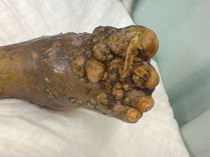
Eumycetoma is a chronic granulomatous[1] fungal disease[2] of humans, affecting mainly the limbs, and sometimes the abdominal and chest walls or the head.[3] Mycetoma pedis (mycetoma of the foot), the most common form of mycetoma, is known widely as the Madura foot. The infection is endemic in Africa, India and Central and South America.[4]
| Mycetoma | |
|---|---|
 | |
| Madura Foot | |
| Specialty | Infectious disease |
Signs and symptoms
The initial lesion is a small subcutaneous swelling following minor trauma. Later, sinuses that discharge purulent and seropurulent exudates containing grains which are fungal colonies are formed.[3][5] Destruction of deeper tissues, and deformity and loss of function in the affected limbs may occur in later stages.
Causes
Mycetoma may be caused by bacteria from the phylum Actinomycetes, or by fungi (Eumycetes) where it is called eumycetoma.[3][5] Bacterial and fungal species that can cause mycetoma are listed below under their characteristic colours of discharge from infected wounds:
Red discharge
- Actinomadura pelletieri (bacterium)
White or yellow discharge
- Acremonium strictum
- Actinomadura madurae (bacterium)
- Aspergillus nidulans
- Noetestudina rosatii
- Phaeoacremonium krajdenii[6]
- Pseudallescheria boydii[7]
Black discharge
- Aspergillus terreus
- Curvularia lunata
- Cladophialophora bantiana
- Exophiala jeanselmei[8]
- Leptosphaeria senegalensis
- Leptosphaeria tompkinsii
- Madurella grisea[9]
- Madurella mycetomatis[10]
- Pyrenochaeta romeroi
Some species of the bacterial genus Nocardia (including Nocardia asteroides and Nocardia brasiliensis) which can cause mycetoma produce a yellow discharge, and those of the bacterial genus Streptomyces (including Streptomyces somaliensis) produce a yellow or red discharge.
Pathogenesis
The disease is usually seen in field workers like farmers, and generally affects men between 20 and 40 years. The disease is acquired by inoculation of grains of fungal spores from the soil through a breach in the skin produced by minor trauma like a thorn prick. The disease then spreads to deeper tissues and also forms sinus tracts leading to skin surface.[5] Mature lesions are characterised by a grainy discharge from these sinuses. These discharges contain fungal colonies and are infective. Spread of infection internally through blood or lymph is uncommon.
Infections that produce a black discharge mainly spread subcutaneously. In the red and yellow varieties deep spread occurs early, infiltrating muscles and bones but sparing nerves and tendons, which are highly resistant to the invasion.[4]
Botryomycosis, also known as bacterial pseudomycosis, produces a similar clinical picture and is caused usually by Staphylococcus aureus.[11] Other bacteria may also cause botryomycosis.[12]
Diagnosis
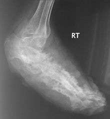
Diagnosis of mycetoma is usually established clinically in endemic areas. X rays and ultrasonography may be employed in evaluating the extent of the disease. X rays findings are extremely variable. The disease is most often observed at an advanced stage that exhibits extensive destruction of all bones of the foot. Rarely, a single lesion may be seen in the tibia where the picture is identical with chronic osteomyelitis. Cytology of fine needle aspirate or pus from the lesion, and tissue biopsy may be undertaken sometimes.[3] Some publications have claimed a "dot in a circle sign" as a characteristic MRI feature for this condition (this feature has also been described on ultrasound).
Differential diagnosis
The following clinical conditions may be considered before diagnosing a patient with mycetoma:
- Tuberculous ulcer
- Kaposi's sarcoma, a vascular tumour of skin usually seen in AIDS.
- Leprosy
- Syphilis
- Malignant neoplasm
- Tropical ulcer[4]
- Botryomycosis,[5] a skin infection usually caused by the bacteria Staphylococcus aureus.
Prevention
No vaccine is available. Simple hygienic precautions like wearing shoes or sandals while working in fields, and washing hands and feet at regular intervals may help prevent the disease.
Treatment
Drugs like ketoconazole,[13] voriconazole,[14] and itraconazole[3] are generally employed in treating the infection. Actinomycetes usually respond well to medical treatment, but the eumycetes are generally resistant and may require surgical interventions including amputation.[5]
Epidemiology
The disease is endemic in tropical and subtropical regions.[3][5] The exact incidence and geographical distribution of mycetoma throughout the world is not known as the disease is usually painless, slowly progressive and presented to health centres only in late stages by majority of patients. Mycetoma has an uneven worldwide distribution.
History
Madura foot or maduromycosis or maduramycosis[15] is described in ancient writings of India as Padavalmika, which, translated means Foot anthill.[5] The first modern description of Madura foot was made in 1842 from Madurai (the city after which the disease was named Madura mycosis) in India, by Gill.[5] The fungal cause of the disease was established in 1860 by Carter.[5]
References
- Motswaledi HM, Mathekga K, Sein PP, Nemutavhanani DL (August 2009). "Paecilomyces lilacinus eumycetoma". Int. J. Dermatol. 48 (8): 858–61. doi:10.1111/j.1365-4632.2008.04047.x. PMID 19659864.
- Brownell I, Pomeranz M, Ma L (2005). "Eumycetoma". Dermatol. Online J. 11 (4): 10. PMID 16403382.
- Davidson's principles and practice of medicine (20th ed.). Churchill Livingstone Elsevier. 2006. p. 373. ISBN 9780443101335.
- Hamilton Bailey's Demonstrations of Physical Signs in Clinical Surgery ISBN 0-7506-0625-8
- Ananthanarayan BA, Jayaram CK, Paniker MD (2006). Textbook of Microbiology (7th ed.). Orient Longman Private Ltd. p. 618. ISBN 978-8125028086.
- Hemashettar BM, Siddaramappa B, Munjunathaswamy BS, et al. (December 2006). "Phaeoacremonium krajdenii, a cause of white grain eumycetoma". J. Clin. Microbiol. 44 (12): 4619–22. doi:10.1128/JCM.01019-06. PMC 1698411. PMID 17005754.
- "Filamentous Fungi".
- Severo LC, Oliveira FM, Vettorato G, Londero AT (March 1999). "Mycetoma caused by Exophiala jeanselmei. Report of a case successfully treated with itraconazole and review of the literature". Rev Iberoam Micol. 16 (1): 57–9. PMID 18473595.
- Vilela R, Duarte OM, Rosa CA, et al. (November 2004). "A case of eumycetoma due to Madurella grisea in northern Brazil" (PDF). Mycopathologia. 158 (4): 415–8. doi:10.1007/s11046-004-2844-y. PMID 15630550.
- Ahmed AO, Desplaces N, Leonard P, et al. (December 2003). "Molecular detection and identification of agents of eumycetoma: detailed report of two cases". J. Clin. Microbiol. 41 (12): 5813–6. doi:10.1128/JCM.41.12.5813-5816.2003. PMC 309011. PMID 14662990.
- "Dorlands Medical Dictionary:botryomycosis". 5 September 2008. Archived from the original on 5 September 2008. Retrieved 10 July 2018.
- "Skin-nontumor Infectious disorders Botryomycosis". PathologyOutlines.com, Inc.
- Capoor MR, Khanna G, Nair D, et al. (April 2007). "Eumycetoma pedis due to Exophiala jeanselmei". Indian J Med Microbiol. 25 (2): 155–7. doi:10.4103/0255-0857.32726. PMID 17582190.
- Loulergue P, Hot A, Dannaoui E, et al. (December 2006). "Successful treatment of black-grain mycetoma with voriconazole". Am. J. Trop. Med. Hyg. 75 (6): 1106–7. doi:10.4269/ajtmh.2006.75.1106. PMID 17172376.
- "Infectious Disorders (Specific Agent) Madura foot/Mycetoma/Maduramycosis". MedTech USA, Inc. Archived from the original on 2014-03-22. Retrieved 2013-08-01.
External links
| Classification | |
|---|---|
| External resources |